C17번 모델
Overview
- 연구 배경: 인간 뇌의 구조와 기능을 이해하기 위한 기본적인 지식 제공을 목표로 하며, 신경 세포의 구성과 뇌의 대규모 조직 구조에 대한 설명을 포함한다.
- 핵심 방법론:
- 신경 세포의 세포체, 수상돌기, 축삭 등의 구조를 기반으로 정보 전달 메커니즘을 설명.
- 전기 신호와 화학 신호의 전달 과정(예: 액션 포텐셜, 시냅스 전달)을 상세히 분석.
- 주요 기여:
- 인간 뇌에 존재하는 860억 개의 신경 세포와 각 세포가 약 1만 개의 다른 신경 세포와 연결되는 사실을 제시.
- 뇌의 10%만 사용한다는 오해를 해소하고, 신경 세포와 신경교세포의 비율이 약 1:1임을 명확히 설명.
- 실험 결과:
- 뇌는 체중의 2%만 차지하지만, 신경 세포 수는 시간에 따라 감소(20세에서 90세까지 85,000개/일 평균 소실).
- 동일한 쌍둥이의 뇌 구조가 유전적 요인보다 환경적 요인에 의해 더 많이 영향을 받는다는 연구 결과 제시.
- 한계점:
- 본 장은 주로 인간 뇌에 초점을 맞추었으나, 다른 종의 뇌 구조에 대한 비교는 다루지 않았으며, 기능적 특성의 지역화에 대한 논의는 제한적임.
📋 목차
대단원 구조
- Chapter 2 Introducing the brain — 뇌의 구조와 기능에 대한 전체 개요
- 1.1 Structure and Function of the Neuron — 신경세포의 기본 구조와 신호 전달 메커니즘
- 1.2 Ten Interesting Facts About the Human Brain — 인간 뇌에 대한 10가지 흥미로운 사실
- The Gross Organization of the Brain — 뇌의 거시적 조직 구조
- 2.1 Gray matter, white matter, and cerebrospinal fluid — 회백질, 백질, 뇌척수액의 구성과 역할
- 2.2 A hierarchical view of the central nervous system — 중추신경계의 계층적 구조
- 2.3 Terms of reference and section — 뇌 해부학의 방향 용어와 단면 분류
- The Cerebral Cortex — 대뇌피질의 구조와 분류
- 3.1 Brodmann’s areas — 브로드만 영역에 의한 피질 분류 체계
- The Subcortex — 피질 하부 구조의 기능과 역할
- 4.1 The basal ganglia — 기저핵의 운동 조절과 기술 학습 기능
- 4.2 The limbic system — 변연계의 감정, 기억 관련 기능
- 4.3 The diencephalon — 시상과 시상하부의 감각 중계 및 항상성 조절
- The Midbrain and Hindbrain — 중뇌와 후뇌의 기능
- 5.1 Superior colliculi / Inferior colliculi — 상구와 하구의 감각-운동 통합
- 5.2 Cerebellum — 소뇌의 운동 정밀도와 균형 조절
- 5.3 Pons / Medulla oblongata — 뇌교와 연수의 생명 유지 기능
Chapter 2 Introducing the brain
Summary
이 장은 뉴런의 구조와 신호 전달 원리에서 출발하여, 대뇌피질, 피질하 구조, 중뇌와 후뇌에 이르는 뇌의 거시적 해부학적 조직을 체계적으로 소개한다. 인간 뇌에 초점을 맞추어 신경세포가 어떻게 다양한 신경구조로 구성되는지를 다루며, 이후 장에서 논의할 인지 기능의 신경학적 기반을 확립하는 데 목적이 있다.
It is hard to begin a apter about the brain without waxing lyrical. e brain is the physical organ that makes all our mental life possible. It enables us to read these words, and to consider thoughts that we have never considered before—or even to create thoughts that no human has considered before. is book will scrat the surface of how this is all possible, but the purpose of this apter is more mundane. It offers a basic guide to the structure of the brain, starting from a description of neurons and working up to a description of how these are organized into different neuroanatomical systems. e emphasis is on the human brain rather than the brain of other species.
Structure and Function of the Neuron
Summary
모든 신경세포(neuron)는 세포체(soma), 수상돌기(dendrite), 축삭(axon)의 세 가지 기본 구조로 이루어진다. 수상돌기는 인접 신경세포로부터 정보를 수신하고, 축삭은 정보를 다른 신경세포로 전달하며, 각 신경세포는 다수의 수상돌기를 갖지만 축삭은 하나만 존재한다. 수상돌기와 축삭의 공간적 배열은 신경세포 유형에 따라 다양하게 나타난다.
All neurons have basically the same structure. ey consist of three components: a cell body (or soma), dendrites, and an axon. Although neurons have the same basic structure and function, it is important to note that there are some significant differences between different types of neurons in terms of the spatial arrangements of the dendrites and axon.
e cell body contains the nucleus and other organelles. e nucleus contains the genetic code, and this is involved in protein synthesis (e.g. of certain neurotransmiers). Neurons receive information from other neurons and they make a “decision” about this information (by anging their own activity) that can then be passed on to other neurons. From the cell body, a number of braning structures called dendrites enable communication with other neurons. Dendrites receive information from other neurons in close proximity. e number and structure of the dendritic branes can vary significantly depending on the type of neuron (i.e. where it is to be found in the brain). e axon, by contrast, sends information to other neurons. Ea neuron consists of many dendrites but only a single axon (although the axon may be divided into several branes called collaterals).
시험 팁
수상돌기(dendrite)와 축삭(axon)을 헷갈리는 학생이 많다. **“수신은 수상돌기, 송신은 축삭”**으로 외우자. 수상돌기는 여러 개이고 축삭은 하나뿐이라는 점도 핵심이다. 신호 방향은 항상 수상돌기 → 세포체 → 축삭 → 시냅스 순서이다.
Ten Interesting Facts About the Human Brain
Summary
- 인간 뇌에는 약 860억 개의 신경세포가 존재한다 (Azevedo et al., 2009).
- 각 신경세포는 약 1만 개의 다른 신경세포와 연결된다.
- 만약 모든 뉴런이 서로 연결된다면 뇌의 지름은 12.5마일(맨해튼 섬 길이)에 달할 것이다 → 뉴런은 소수의 이웃 뉴런과만 연결되며, 장거리 연결은 예외적이다 (Nelson & Bower, 1990).
- “뇌의 10%만 사용한다”는 속설은 오해이며 (Beyerstein, 1999), 뉴런과 글리아세포의 비율은 약 1:1이다 (Azevedo et al., 2009).
- 뇌는 체중의 단 2%만을 차지한다.
- 치아이랑(dentate gyrus)에서 신경세포 재생이 확인되었다 — 뉴런이 재생되지 않는다는 기존 통념은 깨졌다 (Gross, 2000).
- 20~90세 사이 하루 평균 85,000개의 피질 신경세포가 소실되며, 이 기간 피질 뉴런의 약 10%가 사라진다 (Pakkenberg & Gundersen, 1997).
- 일란성 쌍둥이도 뇌 구조가 동일하지 않다 — 3차원 피질 이랑 패턴은 유전보다 비유전적(환경) 요인이 주로 결정한다. 단, 뇌 크기 자체는 유전성이 강하다 (Bartley et al., 1997).
- 자폐증 환자는 뇌가 크지만 (Abell et al., 1999), 뇌 크기와 지능 사이에 단순한 관계는 없다 — 뇌의 효율성은 크기와 무관할 수 있다.
- 남성의 뇌가 더 크지만, 여성의 뇌는 더 많이 접혀 있어(folding) 표면적 증가로 크기 차이를 상쇄할 수 있다 (Luders et al., 2004). 피질 뉴런 총수는 성별과 관련되나 키·체중과는 무관하다 (Pakkenberg & Gundersen, 1997).
글리아세포 (Glia)
뉴런을 둘러싸고 지원하는 비신경 세포로, 뇌에서 뉴런과 약 1:1 비율로 존재한다. 조직 수리(tissue repair), 미엘린(myelin) 형성, 영양 공급, 노폐물 제거 등 필수적인 지원 기능을 수행한다. 뉴런처럼 전기 신호를 전달하지는 않지만, 뇌 기능 유지에 없어서는 안 되는 세포이다.
주의
“뇌의 10%만 사용한다”는 속설은 완전한 오해이다. 이 오해의 기원은 뇌 세포 중 뉴런이 10%뿐이라는 (역시 부정확한) 주장에서 비롯되었다. 실제로 뉴런과 글리아세포의 비율은 약 1:1이며, 뇌의 거의 모든 영역이 활성화된다. 시험에서 이 구분이 자주 출제된다.
Key Terms
Neuron
A type of cell that makes up the nervous system and supports, among other things, cognitive function.
신경세포(neuron)는 신경계를 구성하며 인지 기능을 지원하는 기본 세포 유형이다. 전기적, 화학적 신호를 통해 정보를 처리하고 전달한다.
Cell body
Part of the neuron containing the nucleus and other organelles.
세포체(cell body/soma)는 핵과 세포소기관을 포함하며, 유전 정보 저장 및 신경전달물질 합성을 담당하는 신경세포의 중심 구조이다.
Dendrites
Braning structures that carry information from other neurons.
수상돌기(dendrite)는 인접 신경세포로부터 전기적, 화학적 신호를 수신하는 분기 구조이다. 그 수와 형태는 신경세포 유형에 따라 다양하며, 수신 정보의 양과 복잡성을 결정한다.
Axon
A braning structure that carries information to other neurons and transmits an action potential.
축삭(axon)은 활동전위(action potential)를 전달하여 다른 신경세포로 정보를 보내는 출력 구조이다. 수상돌기가 입력을 담당하는 것과 대비되며, 신경회로 형성의 기초가 된다.
Synapse
e small gap between neurons in whi neurotransmiers are released, permiing signaling between neurons.
시냅스(synapse)는 신경세포 사이의 작은 간극으로, 시냅스전 뉴런의 축삭 말단에서 신경전달물질이 방출되어 시냅스후 뉴런의 수용체에 결합하는 방식으로 정보를 전달한다. 시냅스후 뉴런에서 생성된 시냅스 전위는 수동적으로 전도되며, 여러 수상돌기의 신호가 합산되어 축삭 시작점에서 충분한 강도에 도달하면 활동전위로 전환된다. 수동적 전도는 단거리에 제한되지만, 활성 전도는 활동전위를 통해 장거리 신호 전달을 가능하게 한다.
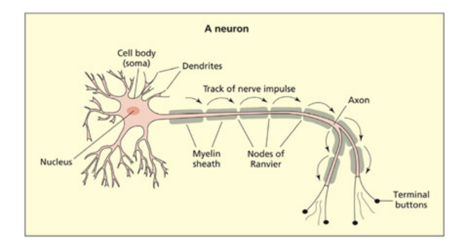
Neurons consist of three basic features: a cell body, dendrites that receive information and axons that send information. In this diagram the axon is myelinated to speed the conduction time.
📊 그림 설명
신경세포(neuron)의 기본 구조를 보여주는 도해이다. 세포체(soma), 수상돌기(dendrite), 축삭(axon)의 세 가지 주요 구성 요소가 표시되어 있다. 축삭은 미엘린(myelin)으로 감싸져 있어 전도 속도가 빨라지는 구조를 나타내며, 정보가 수상돌기에서 수신되어 축삭을 통해 다른 신경세포로 전달되는 방향성을 시각적으로 보여준다.
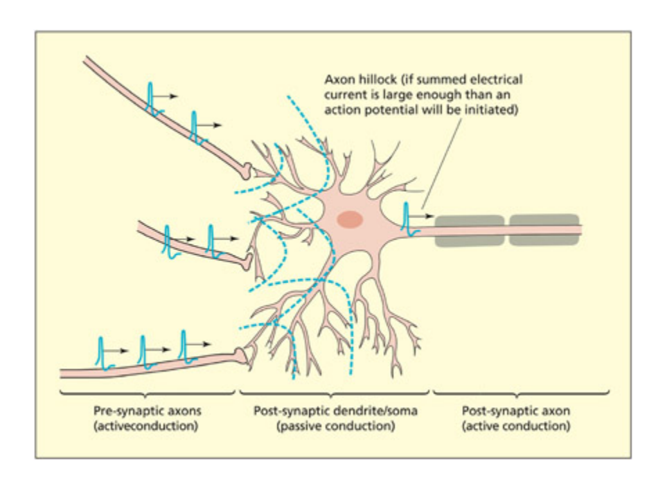
Electrical currents are actively transmied through axons by an action potential. Electrical currents flow passively through dendrites and soma of neurons, but will initiate an action potential if their summed potential is strong enough at the start of the axon (called the hillo).
📊 그림 설명
축삭에서의 능동적 전기 신호 전달(활동전위)과 수상돌기·세포체에서의 수동적 전기 신호 전달을 비교하는 도해이다. 여러 수상돌기로부터 수신된 시냅스 전위가 축삭 시작부(axon hillock)에서 합산되어 충분한 강도에 도달하면 활동전위가 발생하는 과정을 보여준다. 능동적 전도와 수동적 전도의 차이를 시각적으로 구분하여 신경 신호 전달의 핵심 원리를 설명한다.
e terminal of an axon flaens out into a disc-shaped structure. It is here that emical signals enable communication between neurons via a small gap termed a synapse. e two neurons forming the synapse are referred to as presynaptic (before the synapse) and postsynaptic (aer the synapse), reflecting the direction of information flow (from axon to dendrite). When a presynaptic neuron is active, an electrical current (termed an action potential) is propagated down the length of the axon. When the action potential reaes the axon terminal, emicals are released into the synaptic cle. ese emicals are termed neurotransmitters. (Note that a small proportion of synapses, su as retinal gap junctions, signal electrically and not emically.) Neurotransmiers bind to receptors on the dendrites or cell body of the postsynaptic neuron and create a synaptic potential. e synaptic potential is conducted passively (i.e. without creating an action potential) through the dendrites and soma of the postsynaptic neuron. If these passive currents are sufficiently strong when they rea the beginning of the axon in the postsynaptic neuron, then an action potential (an active
electrical current) will be triggered in this neuron. It is important to note that ea postsynaptic neuron sums together many synaptic potentials, whi are generated at many different and distant dendritic sites (in contrast to a simple ain reaction between one neuron and the next). Passive conduction tends to be short range because the electrical signal is impeded by the resistance of the surrounding maer. Active conduction enables longrange signalingsignaling between neurons by the propagation of action potentials.
Key Terms
Action potential
A sudden ange (depolarization and repolarization) in the electrical properties of the neuron membrane in an axon.
활동전위(action potential)는 축삭막에서 발생하는 탈분극과 재분극의 급격한 전기적 변화이다. Na+ 이온의 유입으로 막전위가 역전되고, K+ 이온의 유출로 원래 상태로 복귀하며, 이 과정이 시냅스에서의 신경전달물질 방출로 이어진다.
Neurotransmitters
Chemical signals that are released by one neuron and affect the properties of other neurons.
신경전달물질(neurotransmitter)은 시냅스에서 방출되어 다른 신경세포의 전기적 특성에 영향을 미치는 화학적 신호이다. 축삭 말단에서 방출되어 시냅스후 뉴런의 수용체에 결합함으로써 신호 전달을 매개한다.
Electrical signaling and the action potential
Summary
축삭막의 전압 의존성 이온 채널이 Na+와 K+ 이온의 흐름을 조절하여 활동전위를 생성한다. 휴지 전위는 -70mV이며, 탈분극-재분극-과분극의 단계를 거친다. 미엘린(myelin)이 축삭을 감싸면 랑비에 결절을 통한 도약전도로 전도 속도가 증가하며, 미엘린 파괴는 다발성 경화증 등의 질환과 관련된다.
Ea neuron is surrounded by a cell membrane that acts as a barrier to the passage of certain emicals. Within the membrane, certain protein molecules act as gatekeepers and allow particular emicals in and out under certain conditions. ese emicals consist, among others, of arged
sodium (Na ) and potassium (K ) ions. The balance between these ions on the inside and outside of the membrane is such that there is normally a resting potential of -70 mV across the membrane (the inside being negative relative to the outside).
Voltage-gated ion channels are of particular importance in the generation of an action potential. They are found only in axons, which is why only the axon is capable of producing action potentials. The sequence of events is as follows:
-
- If a passive current of sufficient strength flows across the axon membrane, this begins to open the voltage-gated Na+ channels.
-
- When the channel is opened, then Na+ may enter the cell and the negative potential normally found on the inside is reduced (the cell is said to depolarize). At about –50 mV, the cell membrane becomes completely permeable and the charge on the inside of the cell momentarily reverses. This sudden depolarization and subsequent repolarization in electrical charge across the membrane is the action potential.
-
- The negative potential of the cell is restored via the outward flow of K+ through voltage-gated K+ channels and closing of the voltage-gated Na+ channels.
-
- There is a brief period in which hyperpolarization occurs (the inside is more negative than at rest). This makes it more difficult for the axon to depolarize straight away and prevents the action potential from traveling backwards.
An action potential in one part of the axon opens adjacent voltage-sensitive channels, and so the action potential moves progressively down the length of the axon, starting from the cell body and ending at the axon terminal. The conduction of the action potential along the axon may be speeded up if the axon is myelinated. Myelin is a fatty substance that is deposited around the axon of some cells (especially those that carry motor signals). It blocks the normal transfer and so the action potential
jumps, via passive conduction, down the length of the axon at the points at whi the myelin is absent (called nodes of Ranvier). Destruction of myelin is found in a number of pathologies, notably multiple sclerosis.
임상 사례
다발성 경화증(Multiple Sclerosis)은 미엘린이 파괴되는 대표적 질환이다. 미엘린이 손상되면 도약전도(saltatory conduction)가 불가능해져 신호 전달 속도가 급격히 느려지고, 감각 이상·근력 저하·시력 장애 등이 나타난다. 미엘린의 기능을 이해하면 이 질환의 증상이 왜 발생하는지 논리적으로 설명할 수 있다.
Key Terms
Myelin
A fay substance that is deposited around the axon of some neurons that speeds conduction.
미엘린(myelin)은 일부 신경세포의 축삭 주위에 축적되는 지방성 물질로, 활동전위의 전도 속도를 높이고 에너지 소비를 줄여 신경계의 효율적 신호 전달을 가능하게 한다. 축삭을 감싸는 절연 구조인 수초(myelin sheath)는 이 미엘린으로 구성되어 있다.
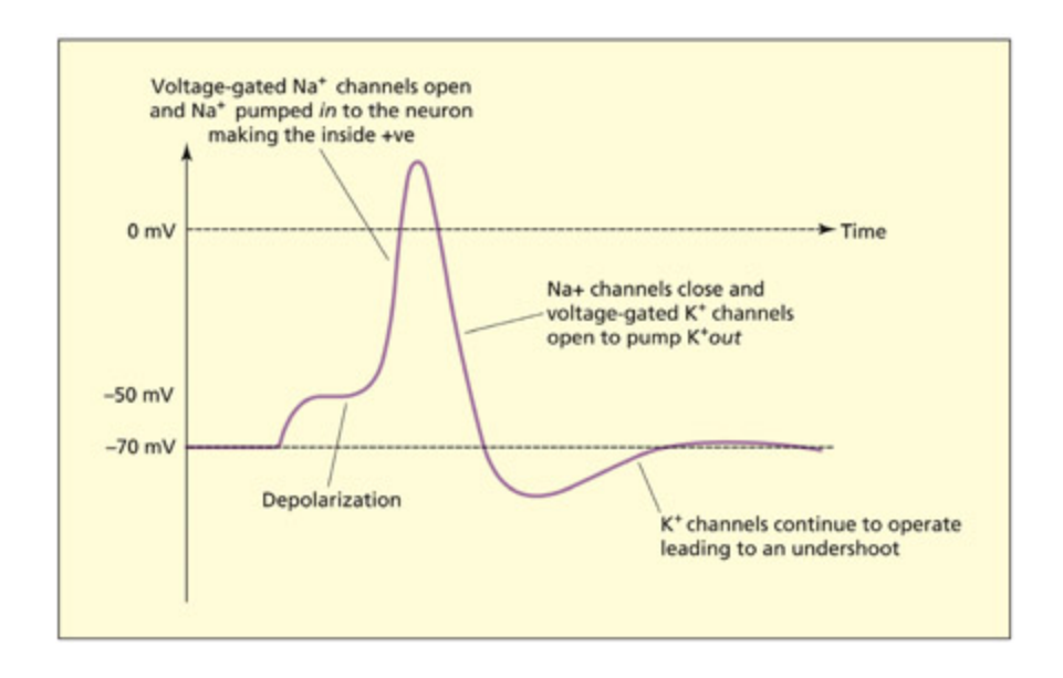
e action potential consists of a number of phases.
📊 그림 설명 — 활동전위 단계별 진행
활동전위의 단계별 진행 과정을 시간에 따른 막전위 변화 그래프로 나타낸 도해이다.
단계 막전위 핵심 기전 ① 휴지 상태 (Resting) -70mV Na⁺/K⁺ 펌프가 Na⁺ 3개 유출, K⁺ 2개 유입 → 안쪽 음전하 유지 ② 역치 도달 (Threshold) ≈ -50mV 수상돌기에서 전달된 수동 전류가 축적되어 역치에 도달 → 전압 의존성 Na⁺ 채널 개방 시작 ③ 탈분극 (Depolarization) -50mV → +30~40mV Na⁺ 채널이 대량 개방, Na⁺ 급속 유입 → 막 안쪽이 양전하로 역전 (그래프의 급상승 구간) ④ 재분극 (Repolarization) +30mV → -70mV Na⁺ 채널 닫힘 + 전압 의존성 K⁺ 채널 개방 → K⁺ 유출로 음전하 회복 (그래프의 급하강 구간) ⑤ 과분극 (Hyperpolarization) -70mV 이하 K⁺ 채널이 약간 늦게 닫혀 K⁺가 초과 유출 → 휴지전위보다 더 음전하 ⑥ 불응기 (Refractory period) → -70mV 복귀 Na⁺/K⁺ 펌프가 이온 분포 원상 복구. 이 기간 동안 재탈분극이 어려워 활동전위의 역방향 전파를 방지 역치에 도달하면 활동전위는 항상 동일한 크기로 발생한다 — 이것이 전부-아니면-전무 법칙(all-or-none principle)이다.
막전위(Membrane Potential) 이해하기
전위(potential)란 두 지점 사이의 전기적 위치 에너지 차이이다. 물이 높은 곳에서 낮은 곳으로 흐르듯, 전하도 전위가 높은 쪽에서 낮은 쪽으로 이동하려는 경향이 있다.
막전위는 세포막을 기준으로 안쪽과 바깥쪽의 전위 차이이며, 관례적으로 막 바깥을 0mV 기준으로 잡고 안쪽을 측정한다. 따라서 -70mV(휴지전위)는 막 안쪽이 바깥보다 70mV만큼 음전하라는 뜻이다.
이 음전하 상태는 Na⁺/K⁺ 펌프가 Na⁺ 3개를 내보내고 K⁺ 2개를 들여보내면서 유지된다. 활동전위 발생 시 Na⁺ 채널이 열려 양이온이 유입되면서(탈분극) 막전위가 급격히 양의 방향으로 올라가는 것이 그래프의 스파이크에 해당한다.
Na⁺/K⁺ 펌프 (Sodium-Potassium Pump)
세포막에 존재하는 능동수송 단백질로, ATP를 소비하여 Na⁺ 3개를 세포 밖으로, K⁺ 2개를 세포 안으로 이동시킨다. 매 cycle마다 양전하 1개가 순(net)으로 빠져나가므로 세포 안쪽이 상대적으로 음전하를 띠게 되며, 이것이 휴지전위(-70mV)를 유지하는 핵심 기전이다.
펌프(Pump) vs 채널(Channel)
펌프 (Pump) 채널 (Channel) 에너지 ATP를 소비하는 능동수송 에너지 불필요, 수동수송 방향 농도 기울기를 거슬러 이온 이동 농도 기울기를 따라 이온 이동 속도 느림 (초당 수백 개) 빠름 (초당 수백만 개) 역할 휴지전위를 유지·복원 활동전위를 생성
- Na⁺/K⁺ 펌프: 평소에 꾸준히 일하면서 안쪽을 -70mV로 유지하는 관리자
- 전압 의존성 이온 채널: 특정 전압에 도달하면 확 열려서 이온을 한꺼번에 통과시키는 수문
활동전위 그래프에서 스파이크가 빠르게 올라갔다 내려오는 건 채널이 열리고 닫히는 것이고, 그 후 원래 상태로 돌아가 유지되는 건 펌프가 담당한다.
Chemical signaling and the postsynaptic neuron
Summary
활동전위가 축삭 말단에 도달하면 신경전달물질이 시냅스 간극으로 방출되어 시냅스후 뉴런의 전달물질-개폐 이온 채널에 결합한다. GABA는 Cl- 채널을 열어 억제 효과를, 아세틸콜린은 흥분 효과를 유발하며, 이러한 시냅스 전위는 수동적으로 전도된다.
When the action potential reaes the axon terminal, the electrical signal initiates a sequence of events leading to the release of neurotransmiers into
the synaptic cleft. Protein receptors in the membrane of the postsynaptic neurons bind to the neurotransmitters. Many of the receptors are transmitter-gated ion channels (not to be confused with voltage-gated ion channels found in the axon). This sets up a localized flow of Na+, K+, or chloride (Cl-), which creates the synaptic potential. Some neurotransmitters (e.g. GABA) have an inhibitory effect on the postsynaptic neuron (i.e. by making it less likely to fire). This can be achieved by making the inside of the neuron more negative than normal and hence harder to depolarize (e.g. by opening transmitter-gated Cl- channels). Other neurotransmitters (e.g. acetylcholine) have excitatory effects on the post-synaptic neuron (i.e. by making it more likely to fire). These synaptic potentials are then passively conducted as already described.
How do neurons code information?
Summary
활동전위의 진폭은 일정하지만, 발화 빈도(spiking rate)가 정보의 코드 역할을 한다. 특정 상황에서만 높은 발화 빈도를 보이는 신경세포들이 공간적으로 그룹화되어 있으며, 이것이 뇌 영역의 기능적 특화의 기초가 된다.
The amplitude of an action potential does not vary, but the number of action potentials propagated per second varies along a continuum. This rate of responding (also called the “spiking rate”) relates to the informational “code” carried by that neuron. For example, some neurons may have a high spiking rate in some situations (e.g. during speech), but not others (e.g. during vision), whereas other neurons would have a complementary profile. Neurons responding to similar types of information tend to be grouped together. This gives rise to the functional specialization of brain regions that was introduced in Chapter 1.
Key Terms
Gray matter
Maer consisting primarily of neuronal cell bodies.
회백질(gray matter)은 주로 신경세포의 세포체로 구성되며, 뇌와 척수에서 정보 처리와 신경 활동의 중심 역할을 한다.
White matter
Tissue of the nervous system consisting primarily of axons and support cells.
백질(white matter)은 미엘린으로 둘러싸인 축삭과 지원 세포(글리아)로 구성되며, 뇌 영역 간 신속한 정보 전달을 담당하는 신경 경로를 형성한다.
Glia
Support cells of the nervous system involved in tissue repair and in the formation of myelin (among other functions).
글리아(glia)는 신경계의 보조 세포로, 조직 수리와 미엘린 형성 등의 지원 기능을 수행한다. 신경세포와 약 1:1 비율로 존재하며, 신경계의 구조적 안정성과 기능적 효율성 유지에 핵심적이다.
Corpus callosum
A large white maer tract that connects the two hemispheres.
뇌량(corpus callosum)은 좌우 두 반구를 연결하는 대규모 백질 경로로, 반구 간 정보 교환과 신경 활동의 동기화를 담당한다.
Ventricles
e hollow ambers of the brain that contain cerebrospinal fluid.
뇌실(ventricles)은 뇌척수액(CSF)으로 채워진 공동 구조이다. 신경세포가 처리하는 정보의 유형은 입력과 출력 경로에 의해 결정되며, 뇌 영역의 기능은 입력 경로를 재구성하면 변화할 수 있다. 따라서 기능의 국재화는 고정적이지 않으며, 입력-출력 상호작용에 의해 결정된다.
If information is carried in the response rate of a neuron, what determines the type of information that the neuron responds to? e type of information that a neuron carries is related to the input it receives and the output it sends to other neurons. For example, the reason neurons in the primary auditory cortex can be considered to carry information about sound is because they receive input from a pathway originating in the colea and
they send information to other neurons involved in more advanced stages of auditory processing (e.g. spee perception). However, imagine that one were to rewire the brain su that the primary auditory cortex was to receive inputs from the retinal pathway rather than the auditory pathway (Sur & Leamey, 2001). In this case, the function of the primary “auditory” cortex would have anged (as would the type of information it carries) even though the region itself was not directly modified (only the inputs to it were modified). is general point is worth bearing in mind when one considers what the function of a given region is. e function of a region is determined by its inputs and outputs. As su, the extent to whi a function can be strictly localized is a moot point.
The Gross Organization of the Brain
Summary
뇌는 회백질(세포체)로 이루어진 대뇌피질, 그 아래의 백질(축삭과 글리아), 중심부의 피질하 구조(기저핵, 변연계, 간뇌)로 구성된다. 백질 경로는 연합로(동일 반구 내), 교련(반구 간, 대표적으로 뇌량), 투사로(피질-피질하 간)로 구분된다. 뇌실은 뇌척수액(CSF)으로 채워져 뇌 보호와 대사 부산물 운반 기능을 수행한다.
Gray matter, white matter, and cerebrospinal fluid
Summary
회백질은 신경세포체로, 백질은 축삭과 글리아로 구성된다. 대뇌피질(회백질) 아래 백질이 위치하고, 그 중심에 피질하 구조(기저핵, 변연계, 간뇌)가 자리한다. 백질 경로는 연합로, 교련(뇌량 포함), 투사로의 세 유형으로 나뉘며, 뇌실의 뇌척수액은 대사물질 운반과 뇌 보호 기능을 수행한다.
Neurons are organized within the brain to form white maer and gray maer. Gray matter consists of neuronal cell bodies. White matter consists of axons and support cells (glia). e brain consists of a highly convoluted folded sheet of gray maer (the cerebral cortex), beneath whi lies the white maer. In the center of the brain, beneath the bulk of the white maer fibers, lies another collection of gray maer structures (the subcortex), whi includes the basal ganglia, the limbic system, and the diencephalon.
White maer tracts may project between different cortical regions within the same hemisphere (called association tracts), may project between different cortical regions in different hemispheres (called commissures; the most important commissure being the corpus callosum) or may project between cortical and subcortical structures (called projection tracts).
e brain also contains a number of hollow ambers termed ventricles. ese were incorrectly revered for 1,500 years as being the seat of mental
life. e ventricles are filled with cerebrospinal fluid (CSF), whi does serve some useful functions, albeit non-cognitive. e CSF carries waste metabolites, transfers some messenger signals, and provides a protective cushion for the brain.
A hierarchical view of the central nervous system
Summary
뇌의 진화는 기존 구조를 대체하지 않고 새로운 구조를 추가하는 방식으로 이루어졌다. 예를 들어 인간의 주요 시각 경로 외에 더 오래된 시각 경로가 공존한다. 뇌실은 4개(좌우 측뇌실, 제3뇌실, 제4뇌실)로 구성되어 뇌척수액이 순환한다.
Brain evolution can be thought of as adding additional structures onto older ones, rather than replacing older structures with newer ones. For example, the main visual pathway in humans travels from the retina to the occipital lobe, but a number of older visual pathways also exist and contribute to vision (see Chapter 6). ese older pathways constitute the dominant form of seeing for other species su as birds and reptiles.
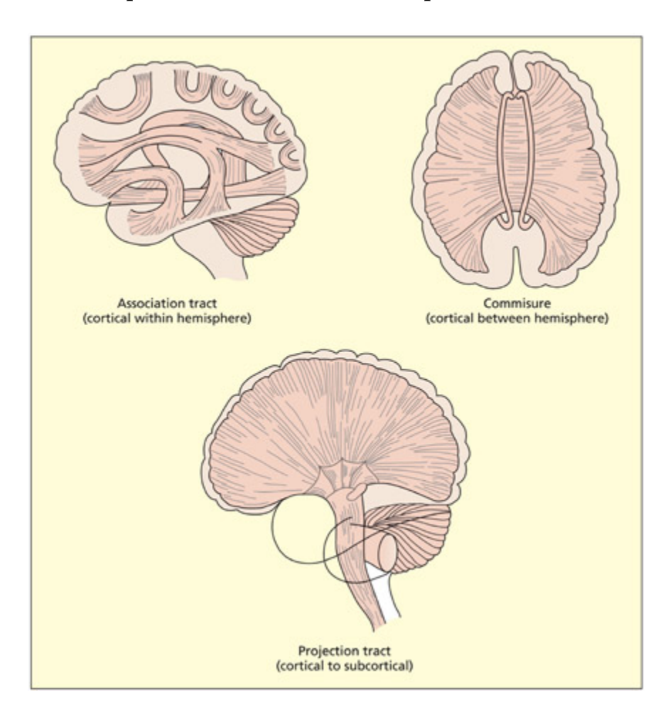
ere are three different kinds of white maer tract, depending on the nature of the regions that are connected.
Adapted from Diamond et al., 1986. © 1986 by Coloring Concepts, Inc. Reprinted by permission of HarperCollins Publishers.
📊 그림 설명
백질 경로의 세 가지 유형을 보여주는 도해이다. 동일 반구 내 피질 영역을 연결하는 연합로(association tracts), 좌우 반구를 연결하는 교련(commissures, 대표적으로 뇌량), 피질과 피질하 구조를 연결하는 투사로(projection tracts)가 각각 구분되어 표시된다. 이 도해는 뇌 영역 간 정보 교환의 구조적 기반을 이해하는 데 도움을 준다.
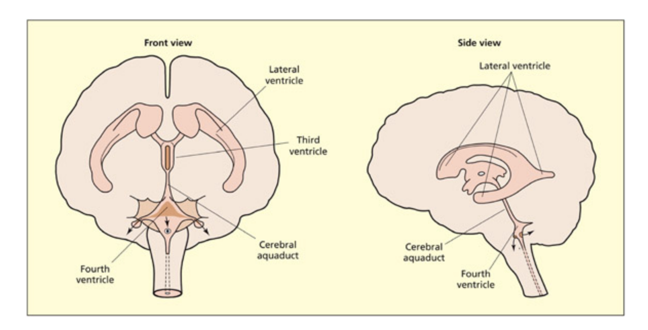
e brain consists of four ventricles filled with cerebrospinal fluid (CSF): the lateral ventricles are found in ea hemisphere, the third ventricle lies centrally around the subcortical structures, and the fourth ventricle lies in the brainstem (hindbrain).
📊 그림 설명
뇌의 4개 뇌실(ventricles) 구조를 보여주는 도해이다. 각 반구에 위치한 좌우 측뇌실(lateral ventricles), 피질하 구조 주변의 제3뇌실(third ventricle), 뇌간(후뇌)에 위치한 제4뇌실(fourth ventricle)이 표시되어 있다. 뇌실은 뇌척수액(CSF)으로 채워져 있으며, 뇌의 보호와 대사 부산물 운반에 기여한다.
Terms of reference and section
Key Terms
Anterior
Towards the front.
전방(anterior/rostral)은 뇌의 앞쪽 방향을 가리키는 해부학적 용어이다.
Posterior
Towards the ba.
후방(posterior/caudal)은 뇌의 뒤쪽 방향을 가리키는 해부학적 용어이다.
Superior
Towards the top.
상방(superior/dorsal)은 뇌의 위쪽 방향을 가리키는 해부학적 용어이다.
Inferior
Towards the boom.
하방(inferior/ventral)은 뇌의 아래쪽 방향을 가리킨다. 뇌의 방향 용어는 전방-후방(anterior-posterior), 상방-하방(superior-inferior), 외측-내측(lateral-medial)의 3차원 좌표 체계로 구성된다.
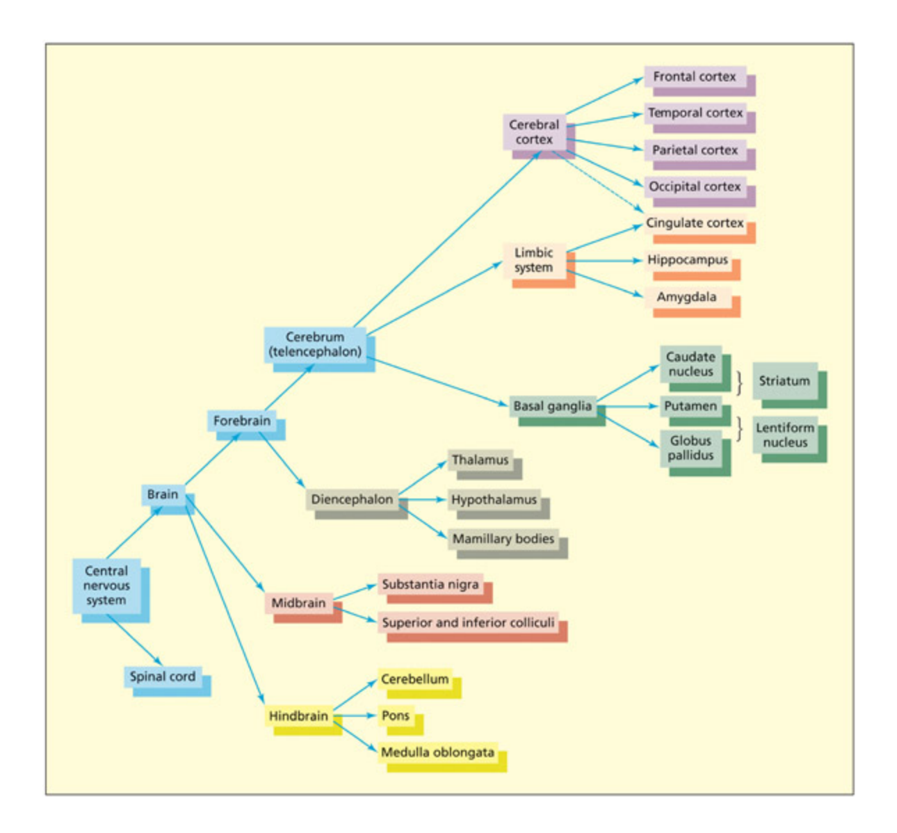
e central nervous system (CNS) is organized hierarically. e upper levels of the hierary, corresponding to the upper branes of this diagram, are the newest structures from an evolutionary perspective.
📊 그림 설명
중추신경계(CNS)의 계층적 조직을 나무 구조(tree diagram)로 나타낸 도해이다. 상위 가지는 진화적으로 가장 새로운 구조(대뇌피질 등)를, 하위 가지는 더 오래된 구조(뇌간 등)를 나타낸다. 이 계층 구조는 뇌 진화가 기존 구조를 대체하지 않고 새로운 구조를 추가하는 방식으로 이루어졌음을 시각적으로 보여준다.
ere are conventional directions for navigating around the brain, just as there is a north, south, east, and west for navigating around maps. Anterior and posterior refer to directions toward the front and the ba of the brain, respectively. ese are also called rostral and caudal, respectively, particularly in other species that have a tail (caudal refers to the tail end). Directions toward the top and the boom are referred to as superior and inferior, respectively; they are also known as dorsal and ventral,
respectively. e terms anterior, posterior, superior, and inferior (or rostral, caudal, dorsal, and ventral) enable navigation in two dimensions: front–ba and top–boom. Needless to say, the brain is three-dimensional and so a further dimension is required. e terms lateral and medial are used to refer to directions toward the outer surface and the center of the brain, respectively; although “medial” is ambiguous, because it is also used in another context. Although it is used to refer to the center of the brain, it is also used to refer to the middle of structures more generally. For example, the medial temporal gyrus lies on the lateral surface of the brain (not the medial surface). It is labeled medial because it lies midway between the superior and inferior temporal gyri.
Key Terms
Dorsal
Towards the top.
배측(dorsal)은 뇌의 윗쪽 방향을 가리키며, superior와 동의어로 사용된다.
Ventral
Towards the boom.
복측(ventral)은 뇌의 아래쪽 방향을 가리키며, inferior와 동의어로 사용된다.
Lateral
e outer part (cf. medial).
외측(lateral)은 뇌의 바깥쪽 표면 방향을 가리키며, 내측(medial)과 대비되는 개념이다.
Medial
In or toward the middle.
내측(medial)은 뇌의 중심부 방향을 가리킨다. 뇌의 단면은 관상면(coronal, 수직-양반구), 시상면(sagittal, 수직-한반구), 수평면(axial)의 세 가지 방식으로 분할된다. 시상면이 반구 사이를 지나면 정중면(midline/medial section)이라 한다.
e brain can be sectioned into two-dimensional slices in a number of ways. A coronal cross-section refers to a slice in the vertical plane through both hemispheres (the brain appears roundish in this section). A sagittal section refers to a slice in the vertical plane going through one of the hemispheres. When the sagial section lies between the hemispheres it is called a midline or medial section. An axial (or horizontal) section is taken in the horizontal plane.
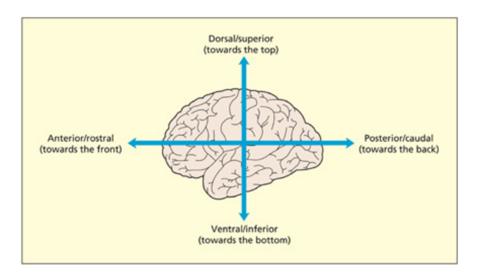
Terms of reference in the brain. Note also the terms lateral (referring to the outer surface of the brain) and medial (referring to the central regions).
📊 그림 설명
뇌의 해부학적 방향 용어를 보여주는 도해이다. 전방(anterior/rostral), 후방(posterior/caudal), 상방(superior/dorsal), 하방(inferior/ventral)의 방향이 뇌 이미지 위에 표시되어 있다. 또한 외측(lateral, 뇌 바깥쪽 표면)과 내측(medial, 뇌 중심부) 방향도 함께 나타나 있어, 뇌 해부학에서 사용되는 3차원 좌표 체계를 한눈에 파악할 수 있다.
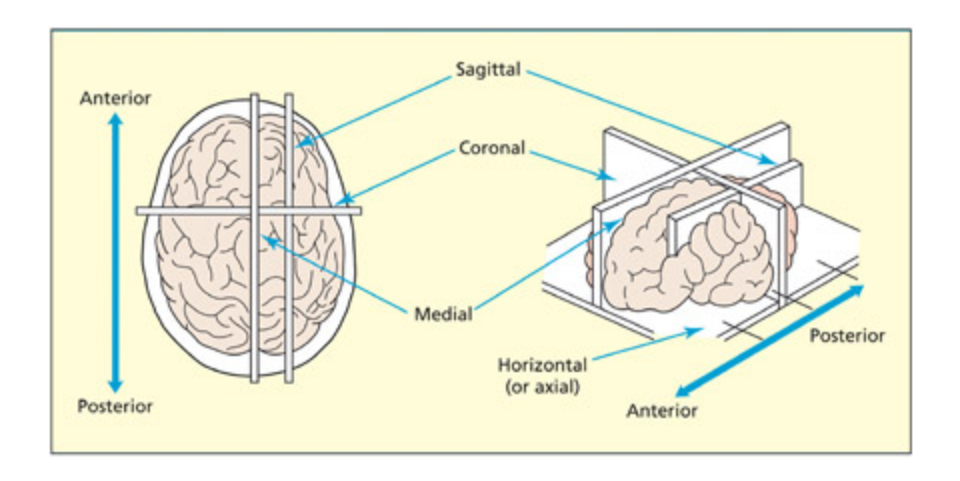
Terms of sections of the brain.
Adapted from Diamond et al., 1986. © 1986 by Coloring Concepts Inc. Reprinted by permission of HarperCollins Publishers.
📊 그림 설명
뇌를 2차원 단면으로 절단하는 세 가지 방법을 보여주는 도해이다. 관상면(coronal, 수직으로 양 반구를 관통), 시상면(sagittal, 수직으로 한 반구를 관통), 수평면(axial/horizontal)이 각각 표시되어 있다. 시상면이 양 반구 사이를 지나가면 정중면(midline/medial section)이라 하며, 이러한 단면 분류는 뇌 영상 분석에서 필수적인 기초 개념이다.
The Cerebral Cortex
Summary
대뇌피질은 좌우 두 반구(2개)로 구성된 주름진 회백질 시트이다. 진화적으로 접힌 구조가 발달하여 표면적 대 부피 비율을 높였으며, 돌출 부분은 이랑(gyri), 함몰 부분은 고랑(sulci)이라 한다.
e cerebral cortex consists of two folded sheets of gray maer organized into two hemispheres (le and right). e surface of the cortex has become increasingly more convoluted with evolutionary development. Having a folded structure permits a high surface area to volume ratio and thereby permits efficient paaging. e raised surfaces of the cortex are termed gyri (or gyrus in the singular). e dips or folds are called sulci (or sulcus in the singular).
Key Terms
Gyri (gyrus = singular)
The raised folds of the cortex.
이랑(gyrus, 복수형 gyri)은 대뇌피질 표면에서 볼록하게 솟아오른 주름이다. 뇌의 표면적을 늘려 제한된 두개골 공간 안에 더 많은 신경세포를 수용할 수 있게 한다.
Sulci (sulcus = singular)
e buried grooves of the cortex.
고랑(sulci)은 피질 표면의 함몰된 주름이다. 피질은 약 3mm 두께로 세포층에 따라 분류되며, 대부분은 6층 구조의 신피질(neocortex)이다. 중간피질(mesocortex, 대상회/섬엽 포함)과 이소피질(allocortex, 일차후각피질/해마 포함)도 존재한다.
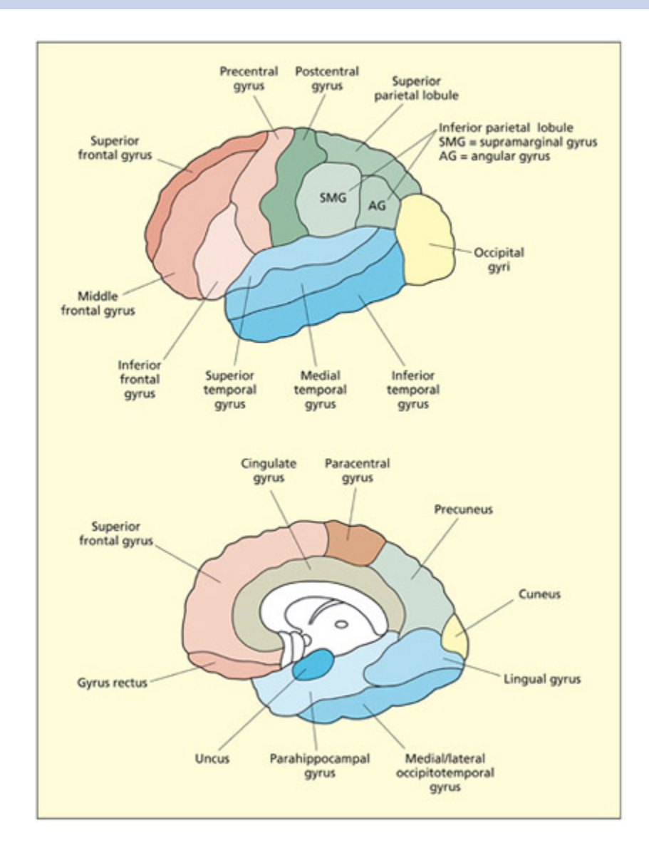
e main gyri of the lateral (top) and medial (boom) surface of the brain. e cortical sulci tend to be labeled according to terms of reference. For example, the superior temporal sulcus lies between the superior and medial temporal gyri.
📊 그림 설명
뇌의 외측면(lateral surface)과 내측면(medial surface)의 주요 이랑(gyri)을 표시한 도해이다. 상단은 외측면에서 보이는 전두엽, 두정엽, 측두엽, 후두엽의 주요 이랑을, 하단은 내측면에서 보이는 대상회(cingulate gyrus) 등의 구조를 보여준다. 고랑(sulci)의 명칭이 방향 용어에 따라 부여되는 규칙(예: 상측두고랑은 상측두이랑과 중측두이랑 사이)을 이해하는 데 도움이 된다.
엽(Lobe) vs 이랑/회(Gyrus) 구분
엽(lobe)과 이랑/회(gyrus)는 서로 다른 해부학적 단위이다.
엽 (Lobe) 이랑/회 (Gyrus) 규모 대분류 — 피질을 크게 나눈 영역 소분류 — 엽 안의 개별 주름(볼록한 부분) 개수 4개 (전두엽·두정엽·측두엽·후두엽) 수십 개 경계 주요 고랑/열(fissure)로 구분 작은 고랑(sulcus)으로 구분 예시 측두엽 (temporal lobe) 상측두회 (superior temporal gyrus) 즉, 엽은 큰 구역, 이랑/회는 그 구역 안의 개별 주름이다. “두정엽”은 뇌의 상부 후방 전체 영역을 가리키고, “각회(angular gyrus)“나 “연상회(supramarginal gyrus)“는 두정엽 안에 있는 특정 이랑을 가리킨다.
한글 용어에서 엽(葉) = lobe, 회(回) = 이랑 = gyrus로 같은 뜻이다.
e cortex is only around 3 mm thi and is organized into different layers that can be seen when viewed in cross-section. e different layers
reflect the grouping of different cell types. Different parts of the cortex have different densities in each of the layers. Most of the cortex contains six main cortical layers, termed the neocortex (meaning “new cortex”). Other cortical regions are the mesocortex (including the cingulate gyrus and insula) and the allocortex (including the primary olfactory cortex and hippocampus).
Key Terms
Brodmann's areas
Regions of cortex defined by the relative distribution of cell types across cortical layers (cytoarchitecture).
브로드만 영역은 피질층의 세포구축(cytoarchitecture)에 따라 정의된 약 52개 영역이다. 피질은 4개 엽으로 나뉘며, 외측열(sylvian fissure)처럼 경계가 명확한 경우와 모호한 경우가 있다. 내측면의 대상피질, 측두엽 아래의 섬엽(insula)도 중요한 구조이다.
Brodmann’s areas
Summary
대뇌피질은 세 가지 기준으로 분류된다: (1) 이랑과 고랑 패턴, (2) 세포구축(cytoarchitecture) 기반의 브로드만 영역(약 52개, BA1~BA52), (3) 기능 기반(일차시각피질=BA17 등). 피질은 전두엽, 두정엽, 측두엽, 후두엽의 4개 엽으로 나뉘며, 고차피질 영역은 기능적 분류가 어렵다.
The lateral surface of the cortex of each hemisphere is divided into four lobes: the frontal, parietal, temporal and occipital lobes. The dividing line between the lobes is sometimes prominent, as is the case between the frontal and temporal lobes (divided by the lateral or sylvian fissure), but in other cases the boundary cannot readily be observed (e.g. between temporal and occipital lobes). Other regions of the cortex are observable only in a medial section, for example the cingulate cortex. Finally, an island of cortex lies buried underneath the temporal lobe; this is called the insula (which literally means “island” in Latin).
There are three different ways in which regions of cerebral cortex may be divided and, hence, labeled:
- Regions divided by the pattern of gyri and sulci. The same pattern of gyri and sulci is found in everyone (although the precise shape and size varies greatly). As such, it is possible to label different regions of the brain accordingly.
-
- Regions divided by cytoarchitecture. One of the most influential ways of dividing up the cerebral cortex is in terms of Brodmann’s areas. Brodmann divided the cortex up into approximately 52 areas (labeled from BA1 to BA52), based on the relative distribution of cell types across cortical layers. Areas are labeled in a circular spiral starting from the middle, like the numbering system of Parisian suburbs. Over the years, the map has been modified.
-
- Regions divided by function. is method tends only to be used for primary sensory and motor areas. For example, Brodmann areas 17 and 6 are also termed the primary visual cortex and the primary motor cortex, respectively. Higher cortical regions are harder (if not impossible) to ascribe unique functions to.
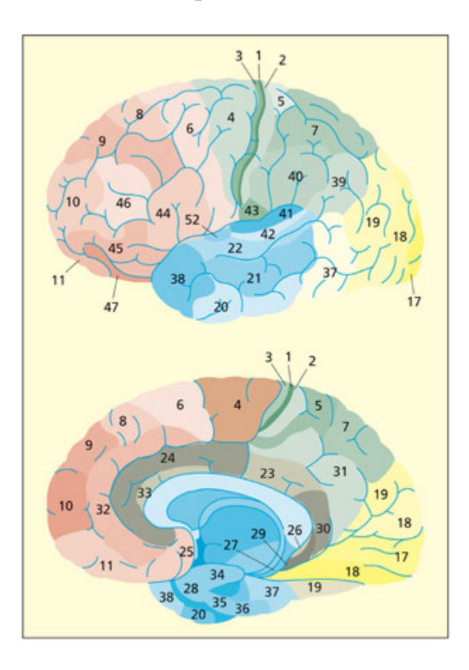
e Brodmann areas of the brain on the lateral (top) and medial (boom) surface.
📊 그림 설명
브로드만 영역(Brodmann areas)을 뇌의 외측면(상단)과 내측면(하단)에 번호와 색상으로 표시한 도해이다. 각 영역은 피질층의 세포구축(cytoarchitecture)에 기반하여 구분되며, BA1~BA52까지 약 52개 영역이 존재한다. 예를 들어 BA17은 일차시각피질, BA6은 일차운동피질에 해당하며, 이 지도는 뇌 기능 연구에서 영역을 지칭하는 표준적 참조 체계로 널리 사용된다.
시험 팁
브로드만 영역 번호를 모두 외울 필요는 없지만, 핵심 영역은 반드시 기억하자: BA17 = 일차시각피질(V1), BA4 = 일차운동피질, BA44/45 = 브로카 영역(언어 산출). 피질 분류의 세 가지 기준(이랑/고랑, 세포구축, 기능)을 구분하는 문제도 자주 출제된다.
The Subcortex
Summary
피질하 구조(subcortex)는 피질과 백질 아래에 위치한 회백질 핵의 집합으로, 기저핵(운동 조절), 변연계(감정/기억), 간뇌(감각 중계/항상성)의 시스템으로 구분된다.
피질하 구조의 위치와 구성
“피질하(subcortical)“란 말 그대로 피질(cortex) 아래라는 뜻이다. 뇌 표면에서 중심부 방향으로 들어가면 다음과 같은 층위를 만난다:
깊이 구성 성질 표면 대뇌피질 (cortex) 회백질 — 신경세포체 층 (약 3mm) 피질 바로 아래 축삭 다발 (신경 배선) 백질 — 미엘린으로 감싸인 축삭이 영역 간 신호를 전달하는 고속도로 더 깊은 중심부 기저핵·시상·편도체·해마 등 다시 회백질 — 신경세포체가 모인 핵(nuclei) 따라서 피질하 구조 = 백질이 아니다. 백질은 피질과 피질하 핵들을 연결하는 배선이며, 시상·기저핵·편도체 같은 핵심 피질하 구조들은 회백질 덩어리(핵) 이다.
Beneath the cortical surface and the intervening white maer lies another collection of gray maer nuclei termed the subcortex. e subcortex is typically divided into a number of different systems with different evolutionary and functional histories.
Key Terms
Basal ganglia
Regions of subcortical gray matter involved in aspects of motor control and skill learning; they consist of structures su as the caudate nucleus, putamen, and globus pallidus.
기저핵(basal ganglia)은 미상핵, 조가비핵, 담창구으로 구성된 피질하 회백질로, 운동 조절과 기술 학습에 관여한다.
Limbic system
A region of subcortex involved in relating the organism to its present and past environment; limbic structures include the amygdala, hippocampus, cingulate cortex, and mamillary bodies.
변연계(limbic system)는 편도체, 해마, 대상피질, 유두체 등으로 구성되며, 유기체와 환경 간의 관계 형성, 감정 반응, 기억 형성에 핵심적 역할을 한다.
Thalamus
A major subcortical relay center; for instance, it is a processing station between all sensory organs (except smell) and the cortex.
시상(thalamus)은 후각을 제외한 모든 감각 정보를 피질로 중계하는 주요 피질하 구조이다. 감각 기관과 피질 사이의 처리 중심지로서 정보의 초기 필터링과 선택적 전달을 수행한다.
Hypothalamus
Consists of a variety of nuclei that are specialized for different functions that are primarily concerned with the body and its regulation.
시상하부(hypothalamus)는 체온, 배고픔, 갈증, 성적 활동, 내분비 기능 조절 등 신체 항상성 유지에 특화된 다양한 핵으로 구성된다.
The basal ganglia
Summary
기저핵은 미상핵, 조가비핵, 창백핵으로 구성되며, 피질 입력을 창백핵에서 시상으로 전달하여 행동의 확률과 강도를 조절한다. 기저핵 장애는 운동저하증(파킨슨병)이나 운동과다증(헌팅턴병)으로 나타나며, 보상 학습과 습관 형성에도 관여한다.
e basal ganglia are large rounded masses that lie in ea hemisphere. ey surround and overhang the thalamus in the center of the brain. ey are involved in regulating motor activity, and the programming and termination of action (see Chapter 8). Disorders of the basal ganglia can be aracterized as hypokinetic (poverty of movement) or hyperkinetic (excess of movement). Examples of these include Parkinson’s and Huntington’s disease, respectively (see Chapter 8). e basal ganglia are also implicated in the learning of rewards, skills, and habits (see Chapters 9 and 15). e main structures comprising the basal ganglia are: the caudate nucleus (an elongated tail-like structure), the putamen (lying more laterally) and the globus pallidus (lying more medially). e caudate and putamen funnel cortical inputs into the globus pallidus, from whi fibers rea into the thalamus. Different circuits passing through these regions either increase or decrease the probability and intensity of certain behaviors (e.g. voluntary movements).
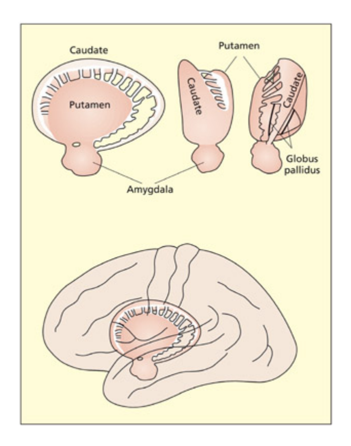
e basal ganglia are involved in motor programming and skill learning.
📊 그림 설명
기저핵(basal ganglia)의 주요 구성 요소인 미상핵(caudate nucleus), 조가비핵(putamen), 창백핵(globus pallidus)의 위치와 구조를 보여주는 도해이다. 기저핵은 시상을 둘러싸고 있으며, 피질로부터 입력을 받아 창백핵을 거쳐 시상으로 출력을 보내는 회로를 형성한다. 이 구조는 운동 프로그래밍, 기술 학습, 보상 기반 학습에 핵심적인 역할을 한다.
The limbic system
Summary
변연계는 환경과의 관계 형성과 감정 반응에 관여한다. 편도체는 위협 자극 탐지, 해마는 학습과 기억, 대상회는 감정/인지 갈등 탐지를 담당한다. 유두체는 기억 기능과 관련되며, 후각구는 환경 자극 탐지에서 변연계와의 연결이 중요하다.
e limbic system is important for relating the organism to its environment based on current needs and the present situation, and based on previous experience. It is involved in the detection and expression of emotional responses. For example, the amygdala has been implicated in the detection of fearful or threatening stimuli (see Chapter 15), and parts of the cingulate gyrus have been implicated in the detection of emotional and cognitive conflicts (see Chapter 14). e hippocampus is particularly important for learning and memory (see Chapter 9). Both the amygdala and hippocampus lie buried in the temporal lobes of ea hemisphere. Other limbic structures are clearly visible on the underside (ventral surface) of the brain. e mamillary bodies are two small round protrusions that have traditionally been implicated in memory (Dusoir et al., 1990). e olfactory bulbs lie on
the under surface of the frontal lobes. eir connections to the limbic system underscore the importance of smell for detecting environmentally salient stimuli (e.g. food, other animals) and its influence on mood and memory.
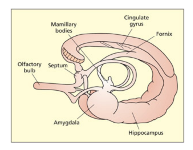
e limbic system.
📊 그림 설명
변연계(limbic system)의 주요 구조를 보여주는 도해이다. 편도체(amygdala), 해마(hippocampus), 대상회(cingulate gyrus), 유두체(mamillary bodies) 등이 표시되어 있다. 변연계는 감정 반응의 탐지와 표현, 학습과 기억, 환경 자극에 대한 적응적 반응에 관여하며, 이들 구조는 측두엽 깊숙이 위치하거나 뇌의 내측면에서 관찰된다.
주의
변연계(limbic system) 구조를 혼동하는 경우가 많다. 편도체(amygdala) = 공포/위협 탐지, 해마(hippocampus) = 학습/기억, 대상회(cingulate gyrus) = 갈등 탐지로 각각의 핵심 기능을 구분하자. 편도체와 해마는 둘 다 측두엽 내부에 위치하지만 기능은 완전히 다르다.
The diencephalon
Summary
간뇌(diencephalon)는 시상과 시상하부로 구성된다. 시상은 외측슬상핵(시각)과 내측슬상핵(청각)을 통해 감각 정보를 일차피질로 중계한다. 시상하부는 체온, 배고픔, 갈증, 내분비 기능 등을 조절하며, 이 부위의 종양은 식이장애, 조기사춘기, 왜소증, 거인증을 유발할 수 있다.
e two main structures that make up the diencephalon are the thalamus and the hypothalamus.
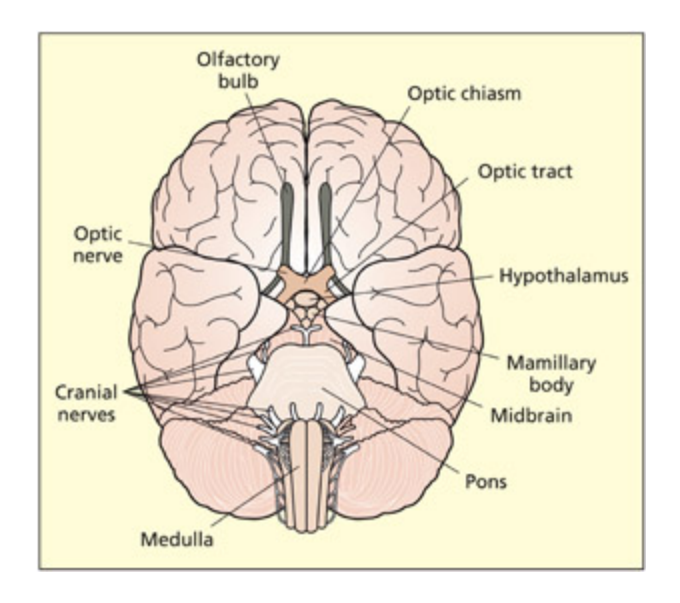
e ventral surface of the brain shows the limbic structures of the olfactory bulbs and mamillary bodies. Other visible structures include the hypothalamus, optic nerves, pons, and medulla.
📊 그림 설명
뇌의 복측면(ventral surface)을 보여주는 도해이다. 변연계 구조인 후각구(olfactory bulbs)와 유두체(mamillary bodies)가 전두엽 아래와 뇌 중앙부에 각각 위치해 있다. 시상하부(hypothalamus), 시신경(optic nerves), 뇌교(pons), 연수(medulla) 등의 구조도 함께 표시되어 있어, 뇌 하면에서 관찰 가능한 주요 해부학적 랜드마크를 종합적으로 보여준다.
e thalamus consists of two interconnected egg-shaped masses that lie in the center of the brain and appear prominent in a medial section. e thalamus is the main sensory relay for all senses (except smell) between the sense organs (eyes, ears, etc.) and the cortex. It also contains projections to almost all parts of the cortex and the basal ganglia. At the posterior end of the thalamus lie the lateral geniculate nucleus and the medial geniculate nucleus. ese are the main sensory relays to the primary visual and primary auditory cortices, respectively.
e hypothalamus lies beneath the thalamus and consists of a variety of nuclei that are specialized for different functions primarily concerned with the body. ese include body temperature, hunger and thirst, sexual activity, and regulation of endocrine functions (e.g. regulating body growth). Tumors in this region can lead to eating and drinking disorders, precocious puberty, dwarfism, and gigantism.
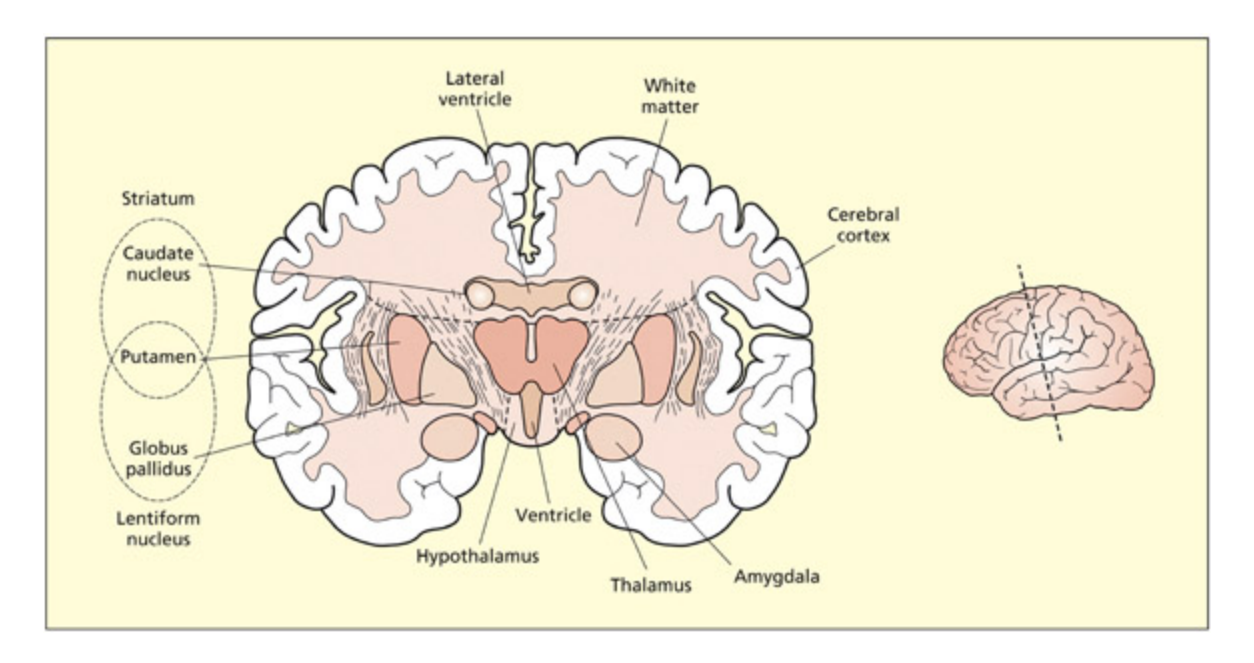
A coronal section through the amygdala and basal ganglia shows the thalamus and hypothalamus as prominent in the midline.
📊 그림 설명
편도체와 기저핵을 지나는 관상면(coronal section) 단면을 보여주는 도해이다. 뇌의 정중선(midline)에서 시상(thalamus)과 시상하부(hypothalamus)가 뚜렷하게 관찰되며, 좌우 대칭적으로 기저핵 구조가 배치되어 있다. 이 단면은 피질하 구조들의 상대적 위치 관계를 이해하는 데 유용하다.
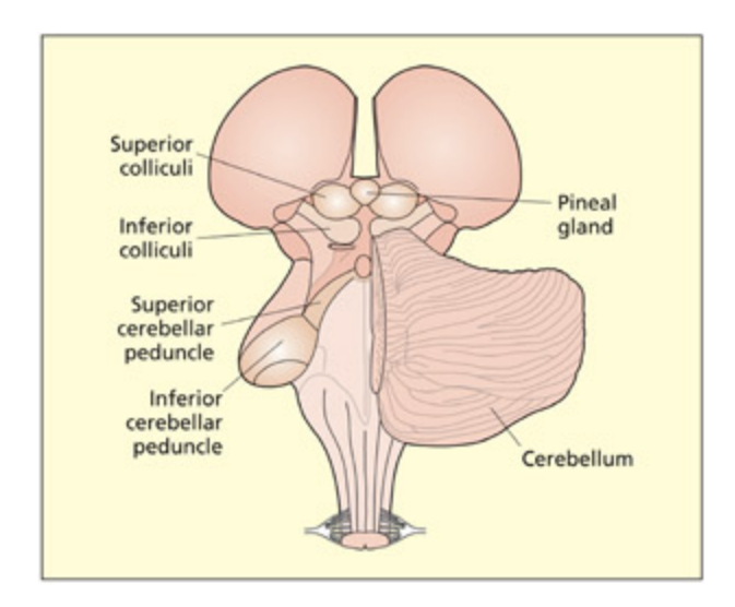
A posterior view of the midbrain and hindbrain. Visible structures include the thalamus, pineal gland, superior colliculi, inferior colliculi, cerebellum, cerebellar peduncle, and medulla oblongata (the pons is not visible but lies on the other side of the cerebellum).
📊 그림 설명
중뇌와 후뇌를 후방(posterior)에서 바라본 도해이다. 시상(thalamus), 송과선(pineal gland), 상구(superior colliculi), 하구(inferior colliculi), 소뇌(cerebellum), 소뇌각(cerebellar peduncle), 연수(medulla oblongata) 등의 구조가 표시되어 있다. 뇌교(pons)는 소뇌 반대편에 위치하여 이 시점에서는 보이지 않으며, 이 도해는 뇌간과 소뇌의 공간적 관계를 파악하는 데 도움을 준다.
임상 사례
시상(thalamus)은 후각을 제외한 모든 감각의 중계소라는 점을 기억하자. 시상하부(hypothalamus) 종양 사례에서는 식이장애, 조기사춘기, 성장 이상 등 다양한 항상성 조절 장애가 나타난다. “시상 = 감각 relay, 시상하부 = 신체 조절”로 구분하면 시험에서 틀리지 않는다.
The Midbrain and Hindbrain
Summary
중뇌와 후뇌는 뇌간의 주요 구성 요소이다. 중뇌는 시각/청각 정보 처리와 운동 반응 조절을, 후뇌는 호흡, 심장 박동, 혈압 등 생명 유지 기능을 담당한다. 소뇌는 운동의 정밀도와 균형을 조절한다.
Key Terms
Superior colliculi
A midbrain nucleus that forms part of a subcortical sensory pathway involved in programming fast eye movements.
상구(superior colliculi)는 중뇌의 피질하 감각 경로로, 빠른 안구 운동의 프로그래밍과 다감각 정보(시각, 청각, 촉각) 통합에 관여한다.
Inferior colliculi
A midbrain nucleus that forms part of a subcortical auditory pathway.
하구(inferior colliculi)는 중뇌의 피질하 청각 경로에 특화된 구조이다. 상구/하구는 진화적으로 오래된 경로로 자극에 대한 신속한 정위 반응을 가능하게 한다. 중뇌의 흑색질(substantia nigra)은 기저핵과 연결되며, 이 부위의 세포 손실은 파킨슨병과 관련된다.
e midbrain region consists of a number of structures, only a few of whi will be considered here. e superior colliculi and inferior colliculi (or colliculus in singular) are gray-maer nuclei. e superior colliculi integrate information from several senses (vision, hearing, and tou), whereas the inferior colliculi are specialized for auditory processing. ese pathways are different from the main x003C;pg>cortical sensory pathways and are evolutionarily older. ey may provide a fast route that enables rapid orienting to stimuli (flashes or bangs) before the stimulus is consciously seen or heard (Sparks, 1999). e midbrain also contains a region called the substantia nigra, whi is connected to the basal ganglia. Cell loss in this region is associated with the symptoms of Parkinson’s disease.
Key Terms
Cerebellum
Structure aaed to the hindbrain; important for dexterity and smooth execution of movement.
소뇌(cerebellum)는 후뇌에 부착된 구조로, 운동 명령과 감각 피드백을 통합하여 운동의 정밀도와 부드러운 실행을 담당한다. 일측 손상은 동측 운동 조화 장애를, 양측 손상은 보행 실조와 구음장애를 유발한다.
Pons
Part of the hindbrain; a key link between the cerebellum and the cerebrum.
뇌교(pons)는 후뇌의 일부로, 소뇌와 대뇌 사이의 핵심 연결 구조이다. 시각 영역으로부터 정보를 받아 안구 운동과 신체 운동을 조절한다.
Medulla oblongata
Part of the hindbrain; it regulates vital functions su as breathing, swallowing, heart rate, and the wake–sleep cycle.
연수(medulla oblongata)는 뇌교에서 돌출되어 척수와 합류하며, 호흡, 삼킴, 심장 박동, 각성-수면 주기 등 생명 유지에 필수적인 기능을 조절한다.
e cerebellum (literally “lile brain”) is aaed to the posterior of the hindbrain via the cerebellar peduncles. It consists of highly convoluted folds of gray maer. It is organized into two interconnected lobes. e cerebellum is important for dexterity and smooth execution of movement. is function may be aieved by integrating motor commands with online sensory feedba about the current state of the action (see Chapter 8). Unilateral lesions to the cerebellum result in poor coordination on the same side of the body as the lesion (i.e. ipsilesional side). Bilateral lesions result in a wide and staggering gait, slurred spee (dysarthria), and eyes moving in a to-and-fro motion (nystagmus). e pons is a key link between the cerebellum and the cerebrum. It receives information from visual areas to control eye and body movements. e medulla oblongata protrudes from the pons and merges with the spinal cord. It regulates vital functions su as breathing, swallowing, heart rate, and the wake–sleep cycle.
Summary and Key Points of the Chapter
Summary
신경세포는 인지 기능의 기초 단위로, 발화 빈도를 통해 정보를 인코딩한다. 회백질(세포체)은 피질의 주름진 구조를, 백질(축삭)은 신경 경로를 형성한다. 피질하 구조에는 운동 조절의 기저핵, 감정/기억의 변연계, 감각 중계의 시상, 항상성 조절의 시상하부가 포함된다.
- e neuron is the basic cell type that supports cognition. Neurons form a densely interconnected network of connections. Axons send signals to other cells and dendrites receive signals.
- Neurons code information in terms of a response rate. ey only respond in certain situations (determined by the input they receive from elsewhere).
- Neurons are grouped together to form gray maer (cell bodies) and white maer (axons and other cells). e cortical surface consists of a folded sheet of gray maer organized into two hemispheres.
ere is another set of gray maer in the subcortex that includes the basal ganglia (important in regulating movement), the limbic system (important for emotion and memory functions) and the diencephalon (the thalamus is a sensory relay center and the hypothalamus is concerned with hemostatic functions).
Example Essay Questions
Summary
이 섹션에서는 신경세포(neuron) 간의 정보 전달 메커니즘을 중심으로, 전기적 신호(action potential)와 화학적 신호(neurotransmitter)의 생성 및 작용 방식을 설명한다. 전기적 신호는 축삭(axon)의 이온 채널(ion channels)을 통해 탈극(depolarization)과 재극(repolarization) 과정을 거쳐 생성되며, 이는 시냅스(synapse)에서 신경전달물질(neurotransmitter)이 수용체(receptor)에 결합하여 postsynaptic neuron로 전달되는 화학적 신호와 연결된다. 또한, 전두엽(forebrain), 중뇌(midbrain), 후뇌(hindbrain)의 기능적 차이를 비교하여, 전두엽은 인지, 감정, 운동 조절을 담당하는 고차원적 기능을, 중뇌는 시각, 청각, 운동 반응의 통합을, 후뇌는 호흡, 심장 박동, 균형 조절 등 자율적 생리 기능을 수행함을 강조한다. 이는 이전 섹션에서 다룬 회백질(gray matter)과 백질(white matter)의 구조적 특성, 기저핵(basal ganglia), 선조절 시스템(limbic system) 등과 연결된 뇌의 계층적 기능 분화(hierarchical functional specialization)를 설명하는 핵심 내용이다.
- How do neurons communicate with ea other?
- Describe how electrical and emical signals are generated by neurons.
- Compare and contrast the different functions of the forebrain, midbrain and hindbrain.
Recommended Further Reading
Summary
이 섹션에서는 신경과학 분야의 핵심 개념을 이해하는 데 도움이 되는 추천 도서를 소개한다. Bear, Connors, Paradiso(2006)의 Neuroscience: Exploring the Brain은 신경과학의 전반적인 내용을 체계적으로 다루며, 신경과학을 중심으로 하는 학위 과정의 학생들에게 적합한 상세한 교과서로, 이 책은 이전 섹션에서 다룬 뉴런의 구조와 기능, 시냅스 전달 메커니즘 등과 같은 내용을 심화 학습할 수 있는 자료로 활용할 수 있다. 반면 Crossman, Neary(2010)의 Neuroanatomy: An illustrated colour text는 복잡한 해부학적 구조를 직관적인 그림과 색상으로 설명해, 초보자에게도 접근성이 높은 간결한 가이드로 추천된다. 또한 Pinel, Edwards(2007)의 A colorful introduction to the anatomy of the human brain은 색칠하기를 통한 적극적인 학습 방식으로, 뇌의 해부학적 구조를 시각적으로 익히는 데 유리한 교재로 소개된다. 이 도서들은 이전 섹션에서 설명된 회백질, 백질, 기저핵 등과 같은 뇌의 거시적 구조 및 기능을 보다 깊이 있게 탐구할 수 있는 자료로 활용될 수 있다.
- Bear, M. F., Connors, B. W., & Paradiso, M. A. (2006). Neuroscience: Exploring the brain (3rd edition). Baltimore, MA: Lippinco Williams & Wilkins. A detailed book that covers all aspects of neuroscience. It is recommended for students whose degree contains significant neuroscience components. e book may be beyond the need of many psyology students.
- Crossman, A. R. & Neary, D. (2010). Neuroanatomy: An illustrated colour text (4th edition). Edinburgh: Harcourt Publishers. A good and clear guide that is not too detailed.
- Pinel, J. P. J. & Edwards, M. (2007). A colorful introduction to the anatomy of the human brain: A brain and psychology coloring book (2nd edition). New York: Pearson. An active way of learning your way around the brain.
퀴즈 : 대뇌 피질, 간뇌 슬라이드
🧠 뇌 구조 요약표
대뇌피질 영역 (Cerebral Cortex)
한글 이름 영어 이름 위치/분류 주요 기능 비고 대뇌피질 Cerebral cortex 뇌 표면 (회백질) 고차 인지 기능 전반 좌우 반구, 주름진 시트 구조 전두엽 Frontal lobe 피질 외측면 전방 인지, 감정, 운동 조절 외측열(sylvian fissure)로 측두엽과 구분 두정엽 Parietal lobe 피질 외측면 상부 감각 통합, 공간 인지 측두엽 Temporal lobe 피질 외측면 하부 청각, 언어, 기억 편도체·해마가 내부에 위치 후두엽 Occipital lobe 피질 외측면 후방 시각 처리 BA17 = 일차시각피질 대상피질 Cingulate cortex 내측면 (중간피질) 감정/인지 갈등 탐지 변연계의 일부, mesocortex 섬엽 Insula 측두엽 아래 매몰 내수용감각, 감정 처리 라틴어 “섬(island)“ 신피질 Neocortex 피질 대부분 고차 인지 기능 6층 구조 중간피질 Mesocortex 대상회, 섬엽 감정/내수용 처리 이소피질 Allocortex 일차후각피질, 해마 후각, 기억 6층 미만 구조 브로드만 영역 Brodmann’s areas 피질 전체 (약 52개) 세포구축 기반 분류 체계 BA1~BA52 일차시각피질 Primary visual cortex 후두엽 (BA17) 시각 정보 처리 일차운동피질 Primary motor cortex 전두엽 (BA6) 수의 운동 제어 일차청각피질 Primary auditory cortex 측두엽 청각 정보 처리 와우로부터 입력 수신 이랑 Gyri (gyrus) 피질 표면 돌출부 — 표면적 증가 고랑 Sulci (sulcus) 피질 표면 함몰부 — 외측열 Sylvian fissure 전두엽-측두엽 경계 — 주요 해부학적 경계
피질하 구조 (Subcortical Structures)
한글 이름 영어 이름 위치/분류 주요 기능 비고 기저핵 Basal ganglia 피질하 (시상 주변) 운동 조절, 기술 학습, 보상 학습 미상핵+조가비핵+창백핵 미상핵 Caudate nucleus 기저핵 피질 입력 수신 꼬리 모양 구조 조가비핵 Putamen 기저핵 (외측) 피질 입력 수신 창백핵 Globus pallidus 기저핵 (내측) 시상으로 출력 전달 변연계 Limbic system 피질하 감정, 기억, 환경 적응 편도체+해마+대상회+유두체 등 편도체 Amygdala 측두엽 내부 (변연계) 위협/공포 자극 탐지 해마 Hippocampus 측두엽 내부 (변연계) 학습과 기억 allocortex 유두체 Mamillary bodies 뇌 복측면 (변연계) 기억 기능 작은 둥근 돌출 구조 후각구 Olfactory bulbs 전두엽 하면 (변연계) 후각 처리 변연계와 밀접한 연결 시상 Thalamus 간뇌 (뇌 중심부) 감각 중계 (후각 제외) 계란형, 좌우 연결 외측슬상핵 Lateral geniculate nucleus 시상 후방 시각 정보 중계 → 일차시각피질 내측슬상핵 Medial geniculate nucleus 시상 후방 청각 정보 중계 → 일차청각피질 시상하부 Hypothalamus 간뇌 (시상 아래) 체온, 배고픔, 갈증, 내분비 조절 종양 시 식이장애/왜소증/거인증 뇌량 Corpus callosum 두 반구 사이 (백질) 반구 간 정보 교환 대표적 교련(commissure) 뇌실 Ventricles 뇌 내부 공동 뇌척수액 저장·순환 측뇌실(2)+제3+제4 = 4개 치아이랑 Dentate gyrus 해마 영역 신경세포 재생 확인 성인 신경재생 부위
중뇌·후뇌 (Midbrain & Hindbrain)
한글 이름 영어 이름 위치/분류 주요 기능 비고 상구 Superior colliculi 중뇌 다감각 통합, 빠른 안구 운동 시각+청각+촉각 통합 하구 Inferior colliculi 중뇌 청각 처리 피질하 청각 경로 흑색질 Substantia nigra 중뇌 기저핵과 연결 세포 손실 → 파킨슨병 소뇌 Cerebellum 후뇌 후방 운동 정밀도, 균형, 조화 ”작은 뇌”, 소뇌각으로 부착 소뇌각 Cerebellar peduncle 후뇌-소뇌 연결부 소뇌와 뇌간 연결 뇌교 Pons 후뇌 소뇌-대뇌 연결, 안구/신체 운동 시각 영역에서 입력 수신 연수 Medulla oblongata 후뇌 (최하부) 호흡, 삼킴, 심장 박동, 각성-수면 척수와 합류 송과선 Pineal gland 중뇌 후방 멜라토닌 분비, 수면 조절 후방 도해에서 관찰 뇌간 Brainstem 중뇌+후뇌 생명 유지 기능 총괄 중뇌+뇌교+연수
뉴런 구조 (Neuron Structure)
한글 이름 영어 이름 분류 주요 기능 비고 신경세포 Neuron 세포 정보 처리·전달의 기본 단위 약 860억 개 (인간) 세포체 Cell body (soma) 뉴런 구성 핵·세포소기관 포함, 단백질 합성 수상돌기 Dendrites 뉴런 구성 (입력) 인접 뉴런으로부터 신호 수신 다수 존재 축삭 Axon 뉴런 구성 (출력) 활동전위 전달 → 다른 뉴런으로 정보 전송 1개, 측부(collateral) 분기 가능 축삭 언덕 Axon hillock 축삭 시작부 시냅스 전위 합산 → 활동전위 발생 여부 결정 시냅스 Synapse 뉴런 간 간극 신경전달물질 방출로 신호 전달 시냅스전/시냅스후 뉴런 활동전위 Action potential 전기 신호 축삭 내 능동적 장거리 신호 전달 탈분극→재분극→과분극 시냅스 전위 Synaptic potential 전기 신호 수상돌기/세포체의 수동적 전도 단거리, 합산 가능 미엘린 Myelin 축삭 피복 물질 전도 속도 증가 (도약전도) 파괴 시 다발성 경화증 랑비에 결절 Nodes of Ranvier 미엘린 비피복 부위 도약전도 발생 지점 신경전달물질 Neurotransmitters 화학 신호 시냅스후 뉴런 활성/억제 GABA(억제), 아세틸콜린(흥분) 등 글리아 Glia 지원 세포 조직 수리, 미엘린 형성 뉴런과 약 1:1 비율 회백질 Gray matter 조직 유형 신경세포체로 구성 → 정보 처리 피질, 피질하 핵 백질 White matter 조직 유형 축삭+글리아로 구성 → 신호 전달 경로 연합로/교련/투사로 뇌척수액 Cerebrospinal fluid (CSF) 체액 대사 부산물 운반, 뇌 보호 뇌실 내 순환