The case for single patient studies
Overview
- 연구 배경: 뇌 손상 환자 연구는 인지 신경과학에서 정상 기능을 이해하는 데 중요한 역할을 하며, 이는 “역공학”적 접근을 통해 손상된 부위의 기능을 추론하는 방식으로 이루어진다. 이는 뇌 손상이 인지 기능을 “분할”하는 방식을 밝히는 데 기여한다.
- 핵심 방법론:
- 단일 사례 연구와 집단 연구의 두 가지 주요 접근법을 비교 분석하며, 단일 사례 연구는 인지 프로세스의 세분화를, 집단 연구는 손상-결함 연관성을 규명하는 데 적합하다고 강조한다.
- **트랜스크리아니얼 자기 자극(TMS)**과 **트랜스크리아니얼 직접 전류 자극(tDCS)**과 같은 비침습적 자극 기법을 활용한 가상 손상 모델을 통해 인지 기능의 시간적 특성과 복원 가능성을 탐구한다.
- 주요 기여:
- 전통적 신경심리학과 인지 신경심리학의 차이를 해소하고, 두 접근법의 장점을 결합한 통합적 연구 전략을 제시한다.
- 분리(dissociation)와 연관(association)의 개념을 통해 인지 기능의 모듈성과 비모듈성에 대한 이론적 토대를 마련한다. 예를 들어, 모음과 자음 처리에 대한 독립적인 신경 자원이 존재함을 보여주는 사례를 분석한다.
- 실험 결과:
- 단일 사례 연구에서 특정 환자가 모음만 쓰기 어려움을 보이는 반면, 다른 환자는 자음만 쓰기 오류를 보이는 등, 단일 분리(single dissociation)와 이중 분리(double dissociation) 사례를 통해 인지 프로세스의 분리성을 입증한다.
- TMS/tDCS 기법은 뇌 기능의 일시적 손상과 강화를 유도함으로써 인지 기능의 기초를 탐구하는 데 효과적인 도구로 입증된다.
- 의의 및 한계:
- 뇌 손상 연구는 인지 기능의 모듈성과 복잡성을 이해하는 데 중요한 통찰을 제공하지만, 단일 사례의 희소성과 작업-자원 오류(task-resource artifact) 또는 작업-요구 오류(task-demand artifact)와 같은 방법론적 한계를 극복해야 한다.
- 이중 분리의 해석은 모듈성 이론에 대한 지나친 의존을 피하고, 연결주의 모델과 같은 비모듈적 체계에서도 가능하다는 점을 강조한다.
📋 목차
대단원 구조
- Ways of Acquiring Brain Damage — 뇌 손상의 주요 원인을 분류하고 각 유형의 병리학적 메커니즘을 설명한다
- 1.1 Neurosurgery — 간질 치료를 위한 신경외과 수술과 분리뇌 수술
- 1.2 Strokes (or cerebrovascular accident; CVA) — 뇌졸중의 유형과 혈관 질환
- 1.3 Traumatic head injuries — 개방성/폐쇄성 외상성 두부 손상
- 1.4 Tumors — 뇌종양의 종류와 신경세포에 미치는 영향
- 1.5 Viral infections — 바이러스 감염에 의한 뇌 손상
- 1.6 Neurodegenerative disorders — 알츠하이머병, 파킨슨병 등 신경퇴행성 질환
- Dissociations and Associations — 분리와 연관 개념을 통해 인지 기능의 독립성과 모듈성을 분석한다
- Single-Case Studies — Caramazza의 세 가지 가정을 중심으로 단일 사례 연구의 이론적 기반을 논의한다
- 3.1 Caramazza’s assumptions for theorizing in cognitive neuropsyology — 분할·투명성·보편성 가정
- 3.2 The case for single-case studies — 단일 사례 연구의 정당성과 일반화 가능성
- Group Studies and Lesion-Deficit Analysis — 환자 그룹화 전략과 손상-결함 분석 방법론을 설명한다
- 4.1 Ways of grouping patients — 증후군·인지 증상·해부학적 손상 기반 분류
- 4.2 Caveats and complications — 구조적 영상의 한계와 신골상학적 오류
- Animal Models in Neuropsyology — 비인간 동물 모델의 방법론과 윤리적 고려사항을 다룬다
- 5.1 Cognitive neuroscience in nonhuman animals. — 손상 실험 방법과 해부학적 연결성 추적
- Transcranial Magnetic Stimulation (TMS) — TMS의 작동 원리, 가상 손상 기법, 기능적 통합 연구를 소개한다
- 6.1 How does TMS work? — 전자기 유도 원리와 코일 설계
- 6.2 The “virtual lesion” — 가상 손상 기법과 맹인의 시각 피질 재편성 연구
- 6.3 Using TMS to study functional integration — 시각 영역 간 경쟁과 기능적 통합
- 6.4 Practical aspects of using TMS — 자극 타이밍, 위치, 제어 조건 설계
- Safety and Ethical Issues in TMS Research — TMS 실험의 안전성 기준과 윤리적 지침을 정리한다
- Transcranial Direct Current Stimulation (tDCS) — tDCS의 원리, 아노달/카토달 자극 효과, 재활 치료 적용을 설명한다
Chapter 5 The lesioned brain
Summary
뇌 손상 환자 연구는 역공학적 접근(reverse engineering)을 통해 손상된 부위의 기능을 추론함으로써 정상 인지 기능을 이해하는 데 기여한다. 전통적 신경심리학은 집단 연구를 통해 뇌 영역-인지 능력 간 관계를 분석하고, 인지 신경심리학은 단일 사례의 손상/보존 패턴으로 인지 구성 요소를 추론하며, 두 접근법은 인지 신경과학의 발전으로 통합되고 있다. 집단 연구는 손상-결함 연관성 규명에, 단일 사례 연구는 인지 과정의 세분화에 적합하며, TMS와 tDCS 같은 비침습적 자극 기법과 동물 모델도 인지 기능 탐구에 활용된다.
Studies of humans who have been unfortunate enough to acquire brain damage have provided a ri source of information for cognitive neuroscientists. e basic premise behind the approa is that, by studying the abnormal, it is possible to gain insights into normal function. is is a form of “reverse engineering,” in whi one aempts to infer the function of a component (or region) by observing what the rest of the cognitive system can and can’t do when that component (or region) is removed. In this way, lesions “carve cognition at its seams” (McCarthy & Warrington, 1990).
Patient-based neuropsyology has tended to take two broad forms. In one tradition, whi I shall call classical neuropsychology, aempts have been made to infer the function of a given brain region by taking patients with lesions to that region and examining their paern of impaired and spared abilities. is type of resear has benefited greatly from the development of imaging methods that enable more accurate lesion localization and quantification. It also provides an important source of constraint on functional imaging data. In the second tradition, whi I shall call cognitive neuropsychology, the paern of spared and impaired abilities in and of themselves has been used to infer the building blos of cognition —irrespective of where they are located in the brain. is approa has been particularly informative for guiding the development of detailed informationprocessing models and provides the cognitive framework that underpins mu imaging resear. e sism between these traditions has run deep. For example, many journals either tacitly or explicitly favor one approa over the other. Moreover, ea tradition has tended to rely on its own methodology, with classical neuropsyology favoring group studies and cognitive neuropsyology favoring single-case studies. e development of cognitive neuroscience has led to something of a reconciliation of these traditions, and this textbook discusses both. e key point that one needs to bear in mind is this: the method one ooses should be appropriate to the question one is asking. It will be argued in this apter that group studies are more appropriate for establishing lesion-deficit associations, whereas single-case studies are particularly helpful for establishing how cognitive processes might be subdivided.
Key Terms
Group studies
In neuropsyology, the performance of different patients is combined to yield a group average.
그룹 연구는 다양한 환자의 수행 데이터를 결합하여 집단 평균을 산출하고, 환자 간 변이성을 통계적으로 평균화하여 공통적인 인지 패턴을 파악하는 방법이다. 이 접근법은 단일 사례 연구와 달리 대규모 집단 수준에서 일반화 가능한 인지 특성을 도출하는 데 유리하다.
Single-case studies
In cognitive neuropsyology, the data from different patients are not combined.
단일 사례 연구는 개별 환자의 데이터를 결합하지 않고 별도로 분석하여, 특정 뇌 손상이 인지 기능에 미치는 세부적 영향을 파악하는 데 초점을 맞춘다. 역공학적 접근과 결합하여 손상된 뇌 부위의 기능을 추론하는 데 유리하지만, 집단 수준의 통계적 일반화가 어렵다는 한계가 있다.
Transcranial magnetic stimulation (TMS)
Non-invasive stimulation of the brain caused by a rapidly anging electrical current in a coil held over the scalp.
TMS(경두개 자기 자극)는 두피 위 코일에서 빠르게 변하는 전류가 생성한 자기장을 통해 뇌의 특정 영역을 비침습적으로 자극하는 기법이다. 뇌 영역의 신경 활성을 직접 조절할 수 있어 인지/운동 기능 분석과 가상 손상 연구에 활용되며, 자연적 뇌 손상 연구를 보완하는 도구로 널리 쓰인다.
Transcranial direct current stimulation (tDCS)
Non-invasive stimulation of the brain caused by passing a weak electrical current through it.
tDCS(경두개 직류 자극)는 약한 전류를 뇌에 전달하여 신경 활동을 조절하는 비침습적 기법이다. TMS와 유사한 가상 손상 접근이 가능하면서도, 인지 기능 향상(아노달 자극)까지 유도할 수 있다는 점에서 차별화되며, 재활 치료와 인지 기능의 뇌 기초 탐구에 중요한 임상적 의의를 갖는다.
Naturally occurring brain lesions are “accidents of nature” that occur because of stroke, tumor, head injury, or other types of brain damage. A complementary approa, that in many ways resembles the logic of the lesion method, involves magnetic stimulation of the intact brain to produce what has been described as “virtual lesions” (e.g. Pascual-Leone et al., 1999). is method is called transcranial magnetic stimulation (TMS). e method makes contact with the literature from the classical neuropsyology tradition with its emphasis on lesion location. However, it can also be used to test information-processing theories of cognition because
it can provide information on the timing of cognitive processes. e method has a number of advantages over traditional lesion methods. A newer method is based on the principle of electrical stimulation and is termed transcranial direct current stimulation (tDCS) (Nitse et al., 2008). Like TMS it can be used to temporarily disrupt cognitive function (a virtual lesion approa). However, it can also be used to boost cognitive function whi has important implications for rehabilitation as well as for exploring the brain basis of cognition.
Ways of Acquiring Brain Damage
Summary
뇌 손상의 주요 원인으로 신경외과 수술, 뇌졸중(혈류 차단에 의한 신경세포 사멸), 외상성 두부 손상(물리적 충격), 뇌종양, 바이러스 감염, 신경퇴행성 질환(알츠하이머병, 파킨슨병 등)이 있다. 각 손상 유형은 서로 다른 병리학적 메커니즘을 가지며, 인지 신경과학에서 정상 기능을 이해하기 위한 역공학적 분석의 기초 자료를 제공한다. 손상 유형의 다양성은 집단 연구와 단일 사례 연구 모두에서 실험 설계와 데이터 해석의 토대가 된다.
Brain damage can be acquired in a number of ways, as summarized below:
Neurosurgery
Summary
신경외과 수술은 심한 간질 환자에서 발작 초점을 제거하거나, 뇌량(corpus callosum)을 절단하는 분리뇌 수술을 통해 발작 확산을 억제하는 데 사용된다. 대표적 사례로 HM은 내측두엽 절제 후 심각한 기억상실을 보였고, 분리뇌 환자는 일상생활에는 큰 영향이 없으나 실험실에서 양반구 간 정보 전달 차단이 관찰된다. 현재 수술은 약물 치료가 불가능한 경우에 한해 수행된다.
Operations are occasionally performed in cases of severe epilepsy in whi the focus of the epileptic seizure is surgically removed. One of the most famous cases in neuropsyology, HM, had dense amnesia aer part of his medial temporal lobe was surgically removed (see Chapter 9). Another surgical procedure formerly used to reduce epileptic seizures spreading across the brain was to sever the fibers of the corpus callosum. is operation was referred to as the split-brain procedure. Patients who have undergone this intervention have only mild impairments in daily living, but the impairments can be observed in laboratory conditions in whi stimuli are presented briefly to ea hemisphere (for a review, see Gazzaniga, 2000). Surgical intervention was also previously common in psyiatric patients (see the discussion on the prefrontal lobotomy in Chapter 14). In general, surgical
procedures are only carried out in the absence of suitable pharmacological treatments.
Strokes (or cerebrovascular accident; CVA)
Summary
뇌졸중(CVA)은 뇌 혈류 차단으로 인해 신경세포의 국소적 또는 전반적 사멸을 유발한다. 출혈성 뇌졸중은 동맥 파열로 뇌압이 상승하며, 색전(embolism)이나 혈전(thrombosis)에 의한 혈관 폐색도 주요 원인이다. 동맥류, 혈관종, 동맥경화 등의 혈관 질환이 뇌졸중 위험을 높인다.
Disruptions to the blood supply of the brain (called strokes or cerebrovascular accidents, CVA) can result in global or local death of neurons. If an artery ruptures, this leads to a hemorrhage and an increase in intracranial pressure (typically relieved by surgery). People born with aneurysms are more susceptible to rupture. ese are localized regions of over-elastic artery that may balloon and rupture. Blood vessels may also become bloed if, for example, a fay clot gets pushed from a large vessel into a smaller one (an embolism) or a stationary clot becomes large enough to blo the vessel (thrombosis). Other vascular disorders include angiomas (tangled and tortuous blood vessels liable to rupture) and arteriosclerosis (hardening of the vessel walls).
Traumatic head injuries
Summary
외상성 두부 손상은 40세 미만에서 가장 흔한 뇌 손상 유형으로, 특히 교통사고로 인한 청장년 남성에게 다발한다. 개방성 손상은 두개골 골절과 함께 국소적 손상을 보이고, 폐쇄성 손상은 뇌가 두개골 내부에서 충돌하며 광범위한 손상과 의식 상실을 동반한다.
Whereas vascular disorders tend to affect older people, traumatic head injuries are the most common form of brain damage in people of less than 40 years of age. ey are particularly common in young men as a result of road traffic accidents. Traumatic head injuries are classified in two ways, “open” or “closed,” depending on whether the skull is fractured. Open head injuries oen have more localized injuries; whereas closed head injuries have more widespread effects (as the brain ricoets in the skull) and oen produce loss of consciousness.
Tumors
Summary
뇌는 자궁 다음으로 종양이 흔한 부위이며, 수막종(meningioma)과 신경교종(glioma) 같은 원발성 종양과 다른 부위에서 전이된 전이성 종양으로 구분된다. 종양은 비정상적 세포 증식으로 인해 신경세포에 압력을 가하여 기능 장애나 세포 사멸을 유발한다.
e brain is the second most common site for tumors (aer the uterus), and brain tumors are oen spread from other parts of the body (these are called metastatic tumors). Tumors are caused when new cells are produced in a poorly regulated manner. Brain tumors are formed from supporting cells su as the meninges and glia (termed “meningioma” and “gliomas,” respectively). Tumors adversely affect the functioning of the brain because the extra cellular material puts pressure on the neurons, disrupting functioning and possibly leading to cell death.
Viral infections
Summary
바이러스 감염은 특정 뇌 세포를 표적으로 삼아 신경세포 손상을 유발한다. 대표적으로 헤르페스 단순 뇌염(HSE), HIV, 크로이츠펠트-야콥병(CJD)이 있으며, 이들은 각각 염증, 신경퇴행, 프리온에 의한 급속한 뇌 조직 파괴를 통해 인지 기능에 중대한 영향을 미친다.
A number of viruses target specific cells in the brain. ese include herpes simplex encephalitis (HSE), human immunodeficiency virus (HIV), and Creutzfeldt-Jakob disease (CJD).
Neurodegenerative disorders
Summary
고령화 사회에서 신경퇴행성 질환의 발생이 증가하고 있으며, 가장 흔한 것은 알츠하이머형 치매(DAT)로 뇌 위축과 기억상실이 초기 증상이다. 파킨슨병, 헌팅턴병, 의미치매, 다발성 경색 치매 등도 주요 질환이며, 이들은 급성 뇌 손상과 달리 점진적인 신경세포 기능 저하를 특징으로 한다.
Most western societies have a large ageing population that will, if anything, continue to get larger and older. In 1900, 4 percent of people were over the age of 65; in 2030, 20 percent of the population is estimated to be over 65. An increase in life expectancy is bringing about an increase in degenerative illnesses that affect the brain. By far the most common is dementia of the Alzheimer type (or DAT). is is associated with atrophy in a number of regions of the brain, with memory loss (amnesia) typically being the earliest noted symptom. Other neurodegenerative diseases include Parkinson’s disease and Huntington’s disease (see Chapter 8), Pi’s disease (oen the medical
diagnosis in cases of semantic dementia), and multi-infarct dementia (caused by many small strokes that can be hard to distinguish from DAT).
Dissociations and Associations
Summary
분리(dissociation)는 뇌 손상이 특정 인지 기능만 선택적으로 손상시키는 현상을 의미하며, 이를 통해 신경회로의 기능적 독립성을 추론할 수 있다. 연관(association)은 여러 증상이 함께 나타나는 패턴으로, 주로 신경해부학적 근접성에 기인한다. 이중 분리는 두 과제가 서로 다른 신경 자원에 의존함을 보여주는 핵심 증거이며, 과제-자원 오류와 과제-요구 오류 같은 대안적 설명을 배제하는 데 활용된다.
Key Terms
Split-brain
A surgical procedure in whi fibers of the corpus callosum are severed.
분리뇌 수술은 뇌량(corpus callosum) 섬유를 절단하여 양반구 간 정보 전달을 차단하는 신경외과적 절차이다. 심한 간질 환자에게 시행되며, 양반구의 기능적 독립성을 실험적으로 탐구하는 데 활용된다.
Strokes
Disruption in the blood supply to the brain; also called cerebrovascular accidents (CVA).
뇌졸중은 뇌 혈류 차단으로 인한 신경세포 손상으로, 허혈성(혈전/색전에 의한 폐색)과 출혈성(동맥 파열)으로 구분된다.
Aneurysm
Over-elastic region of artery that is prone to rupture.
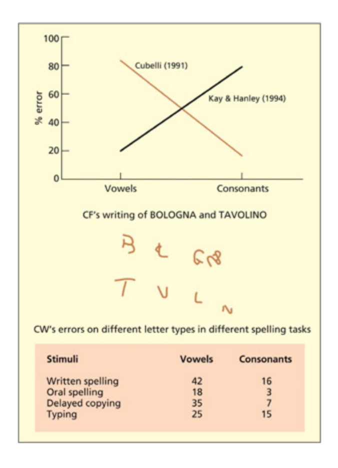
동맥류는 동맥벽이 과도하게 탄력적이어서 파열 위험이 높은 부위이다. 인지 신경심리학적 사례로, Cubelli(1991)의 환자는 모음 쓰기에만 장애를 보였고, Kay와 Hanley(1994)의 환자는 자음 쓰기에만 오류를 보여, 모음과 자음 처리에 별도의 신경 자원이 관여함을 시사하는 단일 분리 및 이중 분리 사례를 제공한다. 다만 과제-자원 오류나 과제-요구 오류 같은 대안적 설명도 고려해야 한다.
In 1990, two very unusual patients came to the aention of Roberto Cubelli (Cubelli, 1991). One patient, CF, was unable to write any vowel leers and le gaps in their place (“Bologna” → B L GN). Another patient, CW, made spelling errors selectively on vowels (e.g. “dietro” → diatro); equivalent errors were not found in his spoken language. By contrast, Kay and Hanley (1994) report a different patient who made spelling errors selectively on consonants (e.g. “record” → recorg). e basic logic behind the cognitive neuropsyological approa is that a difficulty in one domain relative to an absence of difficulty in another domain can be used to infer the independence of these domains. In the case of the patients just discussed, the implication was that the brain has separate neural resources for the processing of wrien vowels relative to consonants. ese neural resources need not lie in different locations of the brain (at least on a millimeter or centimeter scale), but might reflect two different populations of interspersed neurons. Note, also, that it is not clear that one can conclude that the only function of these neurons is the coding of consonants and/or vowels. e difference could be relative and, indeed, without testing a whole range of other stimuli (e.g. digits), it is unwise to conclude exclusivity of function. Nonetheless, it is reasonable to conclude that there are some neural resources predominantly implicated in wrien vowel processing relative to consonants and vice versa.
📊 그림 설명
자음과 모음에 대한 선택적 철자 오류를 보이는 환자 사례를 도식화한 그림이다. 환자 CF는 모음을 쓰지 못하고 빈칸을 남기며, 환자 CW는 모음에서만 철자 오류를 보인다. 이 데이터는 자음과 모음 처리에 별도의 신경 자원이 관여함을 시사하며, 단일 분리 및 이중 분리의 핵심 증거로 활용된다.
Some patients produce spelling errors selectively on either consonants or vowels. is may imply separate neural resources for coding consonants and vowels. Data from Cubelli, 1991.
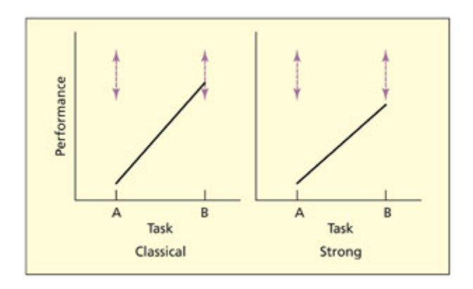
If a patient is impaired on a particular task (task A) but relatively spared on another task (task B), this is referred to as a single dissociation. If the patient performs entirely normally on task B compared with a control group, this has been termed a classical single dissociation, whereas if the patient is impaired on both tasks but is significantly more impaired on one task, this is referred to as a strong single dissociation (Shallice, 1988). In either of these instances, one inference is that task A and task B utilize different cognitive processes with different neural resources. However, other inferences could also be made.
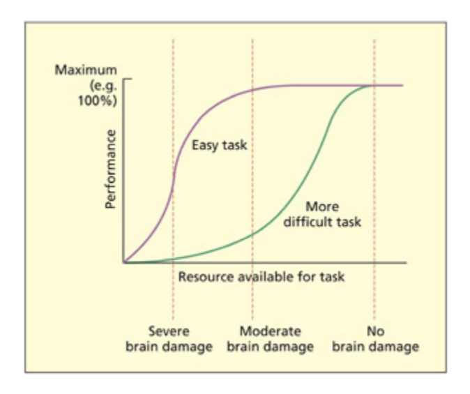
It could be the case that both task A and task B use exactly the same cognitive/neural resources as ea other, but task B requires more of this resource than task A (i.e. task B is harder). If brain damage depletes this
resource, then task B may be relatively or selectively impaired. is has been referred to as a task-resource artifact (Shallice, 1988). Another explanation of a single dissociation is in terms of a task-demand artifact (Shallice, 1988). A task-demand artifact is when a single dissociation occurs because a patient performs one of the tasks suboptimally. For example, the patient may have misunderstood the instructions or have adopted an unusual strategy for performing the task. Task-demand artifacts can be minimized by assessing the patient’s general intellectual functioning, giving clearer instructions or training, using ecologically valid tests, and repeating the same (or similar tests) on several occasions.
Key Terms
Single dissociation
A situation in whi a patient is impaired on a particular task (task A) but relatively spared on another task (task B).
단일 분리는 환자가 과제 A에서 손상을 보이면서도 과제 B에서는 상대적으로 정상 수행을 유지하는 현상으로, 두 과제가 서로 다른 인지/신경 자원을 사용함을 시사한다. 고전적 단일 분리는 과제 B가 통제 범위 내일 때, 강한 단일 분리는 양쪽 모두 손상되었으나 한쪽이 유의미하게 더 심할 때를 말한다.
Task-resource artifact
If two tasks share the same neural/cognitive resource but one task uses it more, then damage to this resource will affect one task more than the other.
과제-자원 오류는 두 과제가 동일한 인지/신경 자원을 공유하되 한 과제가 더 많은 자원을 필요로 할 때, 뇌 손상으로 자원이 감소하면 더 어려운 과제만 선택적으로 손상되는 현상이다. 이는 단일 분리가 반드시 별개의 신경 기반을 의미하지 않을 수 있음을 보여준다.
Task-demand artifact
One task is performed worse than another because the task is performed sub-optimally (but not because some aspect of the task is compromised).
과제-요구 오류는 환자가 지시를 오해하거나 비정상적 전략을 사용하여 한 과제를 비최적적으로 수행함으로써 단일 분리처럼 보이는 현상이다. 이는 과제-자원 오류와 달리 인지 자원 자체의 문제가 아닌 수행 전략의 문제에 기인하며, 명확한 지시, 반복 검사, 생태학적 타당성이 높은 검사로 최소화할 수 있다.
Double dissociation
Two single dissociations that have a complementary profile of abilities.
이중 분리는 환자 1이 과제 A에서 손상/과제 B 보존을 보이고, 환자 2가 반대 패턴을 보이는 현상으로, 두 과제가 서로 다른 신경 자원에 의존함을 강력히 시사한다. 이는 과제-자원 오류를 배제하는 가장 효과적인 방법이며, 인지 기능의 모듈성을 탐구하는 핵심 도구이다.
Dysgraphia
Difficulties in spelling and writing.
실서증(dysgraphia)은 철자 및 글쓰기 능력의 장애로, 뇌졸중이나 신경퇴행성 질환 등으로 인해 발생한다. 모음/자음에 대한 선택적 쓰기 오류 사례는 기능적 분리의 중요한 증거로 활용된다.
Syndrome
A cluster of different symptoms that are believed to be related in some meaningful way.
증후군은 의미 있는 관련성을 가진 증상들의 군집이다. 증상의 연관(association)은 주로 뇌 영역의 해부학적 근접성에 기인하며, 이론적으로는 이중 분리가 더 중요하다. 다만 이중 분리가 반드시 모듈성을 증명하는 것은 아니며, 연결주의 모델 같은 비모듈적 체계에서도 기능적 특화에 의해 이중 분리가 나타날 수 있다. 이중 분리는 유용한 도구이지만 만능이 아니며, 오류 유형 분석과 다양한 관련 과제를 통한 해석이 필수적이다.
In general, almost all neuropsyological studies are aimed at proving that two or more tasks have different cognitive/neural resources and disproving the task-resource and task-demand explanations even if this is not explicitly stated in these terms. In the case of Cubelli’s patients, a task-demand artifact can easily be ruled out because the same task (i.e. writing) was performed in both conditions. One of the most powerful ways of discounting a taskresource artifact is to document a double dissociation, whi merely refers to two single dissociations that have a complementary profile of abilities. To remain with the current example, Kay and Hanley’s patient could write
vowels beer than Cubelli’s patient, whereas Cubelli’s patient could write consonants beer than Kay and Hanley’s.
So far, the discussion has emphasized the importance of dissociations between deficits, but what about associations of deficits? For example, if for every patient that resembled Cubelli’s there were 10, 20, or 100 times as many patients who had comparable dysgraphia for both consonants and vowels, then would this diminish the findings of the dissociation? Some researers would suggest not. ere are some theoretically uninteresting reasons why two symptoms may associate together, the main reason being that they are close together in the brain and so tend to be similarly affected by strokes (or whatever) in that region. For example, patients with difficulties in recognizing faces oen have difficulties in perceiving colors, but this probably reflects neuroanatomical proximity rather than suggesting a “super-module” that is specialized for both. It is the (double) dissociations between the two that count from a theoretical point of view.
📊 그림 설명
고전적 분리와 강한 분리의 차이를 시각적으로 비교한 다이어그램이다. 고전적 분리에서는 한 과제의 수행이 통제 범위(점선) 안에 있고, 강한 분리에서는 두 과제 모두 통제 범위를 벗어나지만 한쪽이 유의미하게 더 손상된다. 이 구분은 단일 분리의 해석에서 과제-자원 오류를 배제하는 데 중요한 기준을 제공한다.
In a classical dissociation, performance on one task lies within the control range (shown by doed lines). In a strong dissociation, both tasks fall outside the control range, but one task is significantly more impaired than the other.
From Shallice, 1988. © Cambridge University Press. Reproduced with permission.
Needless to say, this particular viewpoint has aracted controversy. It has been argued that it is important to know how common a particular dissociation is in order to rule out that it hasn’t been observed by ance (Robertson et al., 1993). For example, if brain damage affects some wrien
leers more than others in a random fashion, then it would still be possible to find patients who appear to have selective difficulties in writing vowels, but it would be a ance occurrence rather than meaningful dissociation. Other researers have focused more on associations between symptoms (so-called syndromes) rather than dissociations. e use of the double dissociation itself has been subject to criticism (see Dunn & Kirsner, 2003). Some have argued that the use of double dissociation implies an endorsement of the notion of modularity (e.g. as specified by Fodor, 1983; see Chapter 1). However, it need not. Shallice (1988) discusses why this argument is wrong by seing up the following thought trap: if modules exist, then double dissociations are a reliable way of uncovering them; double dissociations do exist, therefore modules exist. e way out of this trap, however, is to ask the question: can non-modular systems produce double dis soci ations? It has been demonstrated that other types of cognitive aritecture, su as interactive con nectionist models, can produce double dissociations (Plaut, 1995). e reason why they do so is interesting. It reflects the fact that these systems also contain units that are functionally specialized for certain types of process/ information, even though the system is interactive, and even though these units may respond (to a greater or lesser degree) to a range of stimuli.
📊 그림 설명
과제-자원 오류(task-resource artifact)의 메커니즘을 설명하는 도식이다. 두 과제가 동일한 인지/신경 자원을 공유하되 한 과제가 더 많은 자원을 소비할 때, 뇌 손상으로 가용 자원이 감소하면 어려운 과제만 선택적으로 손상되는 현상을 보여준다. 이는 단일 분리가 반드시 별개의 신경 기반을 의미하지 않을 수 있음을 시각적으로 설명한다.
A task-resource artifact can arise because one task uses more of a cognitive/neural resource than the other (i.e. one task is harder). One could construe brain damage as depleting the amount of resource available. In this instance, at moderate brain damage the patient can still perform the easy task normally. A single dissociation need not reflect different cognitive/neural substrates for the tasks. Adapted from Shallice, 1988.
시험 팁
단일 분리(single dissociation)와 이중 분리(double dissociation)를 구분하는 가장 쉬운 방법: 단일 분리는 “환자 1명, 과제 2개”이고, 이중 분리는 “환자 2명, 과제 2개의 교차 패턴”이다. 핵심은 이중 분리만이 과제-자원 오류(task-resource artifact)를 배제할 수 있다는 것이다. 단일 분리에서 “과제 A만 손상”이 관찰되면, 단순히 A가 더 어려운 과제여서 자원 부족 시 먼저 실패한 것일 수 있다. 이중 분리는 반대 패턴의 환자가 있으므로 이 설명이 불가능해진다.
Some have argued that the reliance on double dissociations is flawed because it requires the study of “pure” cases (Dunn & Kirsner, 2003). However, it need not (Shallice, 1979). First of all, one must be careful to state what is meant by a pure case. For example, imagine that the dysgraphic patients mentioned above also had amnesia. Would the fact that they were not “pure dysgraphic” exclude them from study? is might depend on the theoretical stance one adopts. If one’s theoretical model assumes that writing and memory are independent (as most do), then studying writing in isolation is entirely feasible.
It is worth stating that finding a double dissociation between two patients on two tasks is only part of the neuropsyologist’s toolkit. To interpret their spared and impaired performance, one requires evidence from a range of other relevant tasks. For example, to fully interpret the dysgraphic patients’ impairments it would be interesting to know if they could copy vowels and consonants, or recognize them visually. e types of error that patients produce can also be an important source of information, irrespective of their performance level (i.e. how good or bad they are). For example, the independence of consonants and vowels was initially inferred from the types of errors made in dysgraphia (Caramazza & Miceli, 1990) and not from the double dissociation logic. e double dissociation is useful, but it is not a panacea.
주의
연관(association)과 분리(dissociation)를 혼동하지 말자. 연관은 여러 증상이 함께 나타나는 것이고, 분리는 한 기능만 선택적으로 손상되는 것이다. 시험에서 자주 틀리는 포인트: 연관이 반드시 기능적 관련성을 의미하지 않는다. 얼굴 인식 장애와 색채 지각 장애가 함께 나타나는 이유는 두 기능이 하나의 “슈퍼 모듈”에 속해서가 아니라, 관련 뇌 영역이 해부학적으로 가까이 있어 동일 뇌졸중에 의해 함께 손상되기 때문이다.
Single-Case Studies
Summary
단일 사례 연구의 이론적 기반은 Caramazza가 제시한 세 가지 가정에 의존한다. 분할 가정(fractionation)은 뇌 손상이 인지 모델 내 특정 구성 요소만 선택적으로 손상시킬 수 있다는 것이고, 투명성 가정(transparency)은 손상이 기존 시스템의 구성 요소를 제거하되 새로운 시스템을 생성하지 않는다는 것이며, 보편성 가정(universality)은 모든 인지 시스템이 본질적으로 동일하다는 것이다. 이 가정들은 개별 환자의 비정상 상태를 통해 정상 인지 기능을 추론하는 근거를 제공한다.
Caramazza’s assumptions for theorizing in cognitive neuropsyology
Summary
Caramazza의 세 가지 가정은 인지 신경심리학의 이론적 토대를 형성한다. 분할 가정은 선택적 인지 손상이 가능함을, 투명성 가정은 손상이 기존 시스템을 변형하되 새로운 체계를 만들지 않음을, 보편성 가정은 모든 인지 시스템이 본질적으로 동일함을 전제한다.
Although the use of single cases of brain-damaged individuals to study normal cognitive/brain function began in the mid-nineteenth century, aempts to formalize the logic of this approa were laing for many years. Caramazza provided one of the first serious aempts to do so in the 1980s (Caramazza, 1986, 1992; Caramazza & Badeer, 1989; Caramazza & McCloskey, 1988; McCloskey & Caramazza, 1988).He suggested that three underlying, and unstated, assumptions underpinned almost all neuropsyological studies to date:
-
- The fractionation assumption. e first assumption is that damage to the brain can produce selective cognitive lesions. Note that the assumption is stated with reference to a lesion within a particular cognitive model and not to a lesion to a particular region of the brain (although the two may, of course, be correlated). Caramazza’s arguments were concerned with using observations of braindamaged individuals to inform theories of cognition (cognitive neuropsyology), not to localize cognitive processes in the brain (classical neuropsyology).
-
- The transparency assumption. e transparency assumption states that lesions affect one or more components within the preexisting cognitive system, but they do not result in a completely new cognitive system being created. is assumption is needed because one wishes to study the abnormal in order to understand the normal, and not just to study the abnormal as an end in itself.
-
- The universality assumption. e universality assumption is that all cognitive systems are basically identical.
Key Terms
Transparency assumption
Lesions affect one or more components within the preexisting cognitive system but do not result in a completely new cognitive system being created.
투명성 가정은 뇌 손상이 기존 인지 시스템의 구성 요소를 제거하되 새로운 시스템을 생성하지 않는다는 것이다. 뇌 가소성이나 회복 사례가 이 가정에 도전할 수 있으나, 실어증 후 언어 회복은 기존 모델의 재활성화일 수 있어 반드시 위반은 아니다. 이 가정은 성인기 손상에서 더 잘 유지되며, 손상 직후보다 인지 프로필이 안정화된 시점에서 더 타당하다. 보편성 가정의 문제(개인 차이)는 신경심리학만의 한계가 아니라 인지 신경과학 전체의 과제이며, 단일 사례 연구에서의 개별 차이는 이론적으로 중요한 발견을 포착하는 강점이 될 수 있다.
Caramazza anowledges that these assumptions may, under some situations, not hold true. It is a maer for empirical resear to determine the extent to whi they are true and, hence, the validity of any inference that can be drawn from the study of brain-damaged individuals. Critics have pointed to a number of potential difficulties with the assumptions. Kosslyn and van Kleek (1990) have suggested that whether selective cognitive impairments will be observed (the fractionation assumption) depends on the neural aritecture. For example, selective deficits may be more likely if neurons performing a given operation are clustered together rather than distributed around the brain, and if the neurons are dedicated to one operation rather than shared by many operations. Nevertheless, selective cognitive impairments can be observed and so the fractionation assumption appears to hold true at one level, even if there are some cognitive processes that may be hard to uncover by the lesion method by virtue of an atypical neural aritecture.
e transparency assumption is potentially the most problematic. Basically, one needs to assume that brain damage removes one component of cognition, but does not create, from scrat, a rearranged or different cognitive system. Examples of brain plasticity, and rehabilitation and recovery aer brain damage, might at first appear to be convincing arguments against transparency. But they need not be. For example, imagine that a patient has severe problems in speaking aer a stroke (i.e. aphasia) but that these problems ameliorate over time. is could be taken as prima facie evidence that the brain has somehow reorganized itself aer the stroke. However, it could be that the preexisting cognitive model has just been reinstated rather than that a whole new way of performing the task has been
created. As su, this would not be a violation of the transparency assumption. Plasticity at a neural level is a pervasive aspect of brain function (see also Chapter 9), and need not imply behavioral ange or functional ange. It is important to point out that the assumption is more likely to hold true for brain damage acquired during adulthood than ildhood (omas & Karmiloff-Smith, 2002). It is also worth pointing out that the transparency assumption refers to the cognitive organization of the cognitive system and not necessarily its location. Consider the case of an epileptic ild who has his le hemisphere removed and then learns to speak using the right hemisphere (Vargha-Khadem et al., 1997a). Is that a violation of the transparency assumption? It could be, but it need not be. It depends on whether the new right hemisphere system is cognitively equivalent to the one in the le. e transparency assumption refers to the comparability between premorbid and postmorbid cognitive systems, and not on where su systems are located. Although the debate remains open about the validity of this assumption, a good rule of thumb is that the transparency assumption is less likely to be violated in adult relative to ild cases, and when studied soon aer injury relative to later in time (or if the cognitive profile aer injury remains stable over time).
e universality assumption, that all cognitive systems are basically the same, may also be problematic to neuropsyology. But Caramazza has argued that it is equally problematic for other methods within cognitive neuroscience. Basically, one needs to assume that an individual (or individuals) are representative of the population at large in order to make generalizations to normal cognition. Individual differences, su as they are, are aributable to “noise” (e.g. variations in performance related to time) or other factors that may be related to the efficiency of the cognitive system (e.g. expertise) but need not reflect qualitative differences in the way the task is performed. Of course, if there are individual qualitative differences, then this is theoretically interesting. Finding a framework to explore and account for these differences is a allenge for cognitive neuroscience in general, rather than patient-based neuropsyology in particular. Caplan (1988), however, has argued that individual differences are more of a problem for
singlecase studies relative to other methods because this method gives exaggerated importance to exceptional findings. But this could be construed as the strength of this method rather than a weakness—assuming that the individual differences can be ascribed to something of theoretical interest rather than just “noise.”
The case for single-case studies
Summary
Caramazza는 단일 사례 연구가 인지 신경심리학에서 유일하게 수용 가능한 방법이라고 주장했다. 비손상 집단은 참가자 간 차이가 ‘노이즈’로 간주되어 평균화가 가능하지만, 뇌손상 환자는 각기 다른 인지적 손상(L1-Ln)을 가지므로 개별 분석이 필수적이다. 단일 사례로부터 정상 인지 모델로의 일반화는 가능하지만, 한 환자에서 다른 환자로의 일반화는 어렵다. 이론은 단일 사례에서 구축되는 것이 아니라, 여러 단일 사례와 정상 데이터를 통합하여 검증, 수정, 발전된다.
Caramazza and McCloskey (1988) have gone as far as to suggest that the singlecase study is the only acceptable method in cognitive neuropsyology. e titles of the papers debating this position tell a story of their own. e original paper, entitled “e case for single patient studies” (Caramazza & McCloskey, 1988), was interpreted as the case against group studies. A subsequent paper, “e case against the case against group studies” (Zurif et al., 1989), defended group studies on the grounds that “syndromes [i.e. associations of symptoms] are what the world gives us.” is provoked a paper with a particularly amusing title: “Clinical syndromes are not God’s gi to cognitive neuropsyology: A reply to a rebual to an answer to a response to the case against syndrome-based resear” (Caramazza & Badeer, 1991). To understand this heated debate, it is necessary to take a step ba and consider the argument as initially laid out.
Consider first the logic of testing participants in the non-brain-damaged population. One may recruit a sample of participants (S1 to Sn) and make the assumption, valid or not, that they have broadly equivalent cognitive systems (M). One may then conduct an experiment (E), making the further assumption that all participants carry it out in equivalent ways (i.e. no taskdemand artifacts), and derive a set of observations (O1 to On). In this instance, it is argued that it is quite feasible to average the observations of the group because it is assumed that the only difference between the participants is “noise” (i.e. variations in performance over time, differences in speed or ability).
Consider next the situation in whi one wishes to test a group of braindamaged patients (P1 to Pn). As before, it is assumed that ea has (before their lesion) essentially the same cognitive system (M) and that ea is given the same experiment (E) and complies with the experiment in the same way. However, ea patient may have a different lesion to the cognitive system (L1 to Ln) and so difference in observed performance may be aributable to differences in lesion rather than between-patient noise and, as su, averaging across patients is not possible. Determining where the lesion is in the cognitive system can only be determined on the basis of empirical observation of ea case in turn. It is crucial to bear in mind the distinction between a lesion to a cognitive component (whi is relevant to the discussion here) and an anatomical lesion. At present, there is no magic way of working out what the precise cognitive profile of a given patient will be from a structural lesion (except in the most general terms). us, establishing the cognitive impair ment re quires cognitive testing of individual patients.
| population | |||||
|---|---|---|---|---|---|
| Subjects | S 1 | S2 | S 3 | S 4 | Sn |
| Cognitive systemExperiment | ME | ME | ME | ||
| Observations | 0, | 02 | 0, | O 4 | 0, |
| In a brain-damage | d pop | ulation | |||
| In a brain-damageSubjects | d pop | ulationP 2 | P 4 | Pn | |
| P n | |||||
| Subjects Cognitive system Lesion | P 1 | P 2 | P 3 M L 3 | ML 4 | P, |
| Subjects Cognitive system | P 1 | P 2 | P 3 | М | Ρ, |
Caramazza has argued that it is possible to average observations (O1 to On) across different nonbraindamaged participants (S1 to Sn) because they are assumed to have the same cognitive system (M) that performs the experiment (E) in comparable ways. e same logic may not apply to braindamaged patients (P1 to Pn) because ea patient will have a different cognitive lesion (L), whi cannot be known a priori.
From Caramazza & McCloskey, 1988.
What if one were to establish that a group of patients had identical lesions to the same component of the cognitive system, could one then average across the patients? Caramazza has argued that, although legitimate, the study be comes a series of single-case studies, not a group study, and so the unit of interest is still the single case. To establish that they had the same lesion, one would have to carry out the same set of experiments on ea individually. As su, one would not learn any more from averaging the set of patients than could be learned from a single case itself. e objection is not against the idea of testing more than one patient per se, but rather averaging the results of many patients assumed (but not proven) to be equivalent.
📊 그림 설명
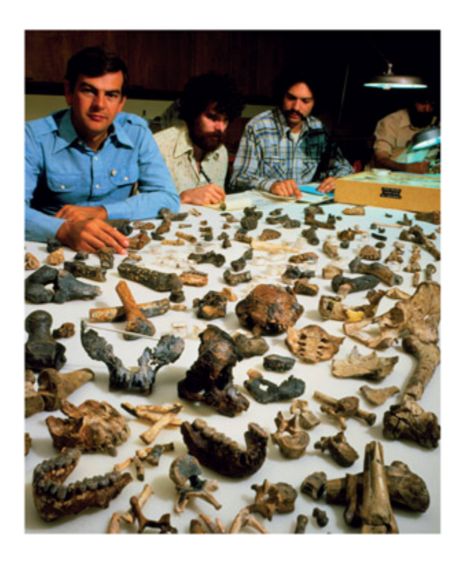
1974년 Donald Johanson이 발견한 318만 년 전 영장류 화석 “Lucy”의 사진이다. Lucy는 직립보행을 했지만 뇌가 작아, 뇌 크기 증가가 직립보행보다 먼저라는 기존 이론을 단일 사례로 반증했다. 이 사례는 단일 사례 연구가 신경심리학에만 국한되지 않으며, 고고학과 인류학에서도 이론 검증의 핵심 방법임을 보여준다.
John Reader/Science Photo Library.
Some of the common objections against the use of the single-case study are that one cannot create a theory based on observations from only a single case, or that it is not possible to generalize from a single case. e counterarguments are that nobody is trying to construct whole new theories of cognition based on a single case. eories, in neuropsyology and elsewhere, must account for a wide range of observations from different sources, both normal and brain-damaged. For example, cognitive models of reading are able to account for different observations found in skilled readers and also account for the different types of acquired dyslexia (drawn from several different single cases). ey must also account for the paern of performance (e.g. the types of error made) as well as the level of performance (i.e. following the logic of dissociations). Although nobody wishes to construct a theory based on a single case, observations from single cases constitute valid data with whi to test, amend, and develop theory. As for the argument that it is not possible to generalize from a single case, the counterquestion would be “generalize to what?.” It is entirely plausible to generalize from a single case to a model of normal cognition. It is, however, mu harder to generalize from one single case to another single case. Two patients with a stroke may have very different cognitive profiles (i.e. one cannot generalize from one case to another), but it should nevertheless be possible for ea particular case to generalize to some aspect of normal cognition.
Evaluation
Summary
단일 사례 연구는 인지 시스템의 구성 요소를 규명하는 데 유효한 방법이지만, 그룹 연구도 다른 유형의 질문에 중요한 역할을 한다. “손상”(lesion)이라는 용어는 인지 모델 내 구성 요소 손상과 생물학적 뇌 손상 두 가지로 해석될 수 있으며, 이는 두 방법론의 상호 보완성을 시사한다.
e argument presented above has emphasized the point that single-case studies are a valid methodology and they may have a particularly important role to play in determining what the components of cognitive systems are. e discussion has also argued that the term “lesion” can be construed both in terms of disruption to a component in a cognitive model, as well as a region of organic brain damage. Does this mean that group studies have no role to play at all? It will be argued that group studies do have an important role to play, and that they may be particularly suited to addressing different types of question from the single-case approa.
시험 팁
집단 연구(group study)와 단일 사례 연구(single-case study)의 역할 구분을 기억하자: 집단 연구는 “어디(where)?”에 답하고(손상 위치-결함 연관), 단일 사례 연구는 “무엇(what)?”에 답한다(인지 구성 요소의 세분화). Caramazza의 핵심 논점은, 뇌 손상 환자는 각각 다른 인지적 손상(L1~Ln)을 가지므로 비손상 집단처럼 단순 평균화가 불가능하다는 것이다. 단, 단일 사례에서 정상 인지 모델로의 일반화는 가능하지만, 한 환자에서 다른 환자로의 일반화는 어렵다.
Group Studies and Lesion-Deficit Analysis
Summary
인지 신경심리학은 인지 기능의 구성 요소에 대한 가설을 제공하고, 전통적 신경심리학은 기능 영상 데이터와의 대비를 통해 뇌 기반 인지 모델을 형성한다. 뇌 손상 환자 연구는 특정 과제에 필수적인 뇌 영역을 식별할 수 있지만, 손상이 대규모이므로 정확한 지역화를 위해 다수의 환자를 고려해야 한다.
e introduction to this apter discussed the historical sism that exists between cognitive neuropsyology, whi is aimed at developing purely cognitive accounts of cognition, and classical neuropsyology, whi is aimed at developing brain-based accounts of cognition. Both approaes fit well within a cognitive neuroscience framework. e cognitive neuropsyology tradition enries the conceptual framework and provides a testable hypothesis about what the likely neural components of cognition are (although not necessarily where they are). e classical neuropsyology tradition provides important contrastive data with functional imaging. ere are several reasons why regions may appear active or inactive in functional imaging tasks, and a region of activity need not imply that a region is critically involved in that particular task. Studies of patients with lesions in that area do enable su conclusions to be drawn. e lesions of patients, however, are typically large and rarely restricted to the region of interest. us, to be able to localize whi region is critical for a given task, several patients may need to be considered.
Ways of grouping patients
Summary
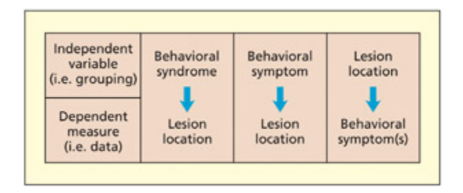
환자 그룹화 전략은 세 가지가 있다: 증후군 기반(증상 패턴으로 분류), 인지 증상 기반(특정 증상을 중심으로 분류), 해부학적 손상 기반(특정 뇌 영역 손상으로 분류). 최근 복셀 단위 병변 매핑(Rorden & Karnath, 2004) 기술로 세밀한 손상 위치 분석이 가능해졌으며, 증상에서 손상 위치로 역추적하는 접근은 여러 뇌 영역의 기여도를 동시에 파악할 수 있다는 장점이 있다.
How does one decide the principle by whi patients should be grouped in order to associate lesion sites with deficits? ere are at least three approaes in the literature:
-
- Grouping by syndrome. Patients are assigned to a particular group on the basis of possessing a cluster of different symptoms. is approa is particularly common in psyiatric studies (e.g. of sizophrenia), but there are equivalent approaes in neuropsyology (e.g. the aphasia subtypes identified by Goodglass and Kaplan, 1972).
-
- Grouping by cognitive symptom. Patients are assigned to a particular group on the basis of possessing one particular symptom (e.g. auditory hallucinations; difficulty in reading nonwords). ey may also possess other symptoms, but, assuming that the other symptoms differ from case to case, the method should be sensitive to the symptom under investigation.
-
- Grouping by anatomical lesion. Patients are selected on the basis of having a lesion to a particular anatomical region. is region may have been identified as interesting by previous functional imaging studies. is method need not require that patients have damage exclusively to the region of interest. e patients may have additional damage elsewhere, but, assuming that the other lesions differ from case to case, the method should be sensitive to the region in question (Damasio & Damasio, 1989).
ere is no right or wrong way of deciding how to group patients, and to some extent it will depend on the precise question being addressed. e method of grouping cases by syndrome is likely to offer a more coarse level of analysis, whereas grouping according to individual symptoms may provide a more fine-grained level of analysis. In general, the syndromebased approa may be more appropriate for understanding the neural correlates of a given disease pathology rather than developing theories concerning the neural basis of cognition.
📊 그림 설명
손상-결함 분석을 위한 세 가지 환자 그룹화 전략을 도식화한 그림이다. 증후군 기반(증상 패턴), 인지 증상 기반(특정 증상 중심), 해부학적 손상 기반(특정 뇌 영역 손상) 분류를 비교하여, 각 접근법의 분석 수준과 적용 맥락의 차이를 시각적으로 보여준다.
ere are at least three different ways of grouping patients to carry out a lesion-deficit analysis.
e method of grouping patients by symptom (2 in the list above) and then finding out what regions of damage they have in common is relatively new. is is made feasible by new teniques that compare the location of lesions from MRI scans of different patients on a voxel-by-voxel basis thus producing a fine-grained statistical map of the likely lesion “hot spot” (Rorden & Karnath, 2004). For example, it has been used to separate out the different contributions of frontal regions in tests of executive function (Shammi & Stuss, 1999; Stuss et al., 2002). One advantage of working forward from a symptom to a lesion location is that it could potentially reveal more than one region as being critically involved. For example, let’s assume that a deficit can arise from damage to either region X or region Y. If one were to initially group patients according to whether they have damage to region X and test for a deficit (3 in the list above), then one could falsely conclude that region X is the key region that gives rise to this deficit and the method would not detect the importance of region Y. e main situation in whi one would group patients by lesion site and then test for the presence of a particular symptom (3 in the list above) is if one has a specific testable prediction about what the region is critical for (e.g. the region has been implicated by functional imaging studies).
Caveats and complications
Summary
손상-결함 분석에는 두 가지 주의점이 있다. 첫째, 구조적 영상 기술은 종양이나 부종 등으로 정확한 손상 범위를 추정하기 어려우며, 신뢰할 수 있는 영상은 발병 3개월 후에 얻는 것이 바람직하다. 둘째, 특정 영역 X의 손상 후 기능 F가 손상되었다고 해서 F가 X에 위치한다고 단정하는 것은 신골상학적 오류이며, X가 F의 일부 측면에 필수적이라는 신중한 결론이 적절하다.
Key Terms
Edema
A swelling of the brain following injury.
뇌 부종(edema)은 손상 후 뇌 조직의 수분 증가로 인해 뇌압이 상승하고 신경세포 기능이 저하되는 현상이다. 종양 주변이나 폐쇄성 두부 손상에서 흔히 관찰되며, 손상 범위를 정확히 추정하기 어렵게 만든다.
Diaschisis
A discrete brain lesion can disrupt the functioning of distant brain regions that are structurally intact.
디아시시스(diaschisis)는 국소적 뇌 손상이 구조적으로 온전한 원거리 뇌 영역의 기능까지 방해하는 현상이다. 예를 들어, 좌측 전두엽 손상이 좌측 하후두측두엽의 활동을 감소시킬 수 있으며, 이는 두 영역이 특정 인지 과제에서 협력적으로 작동하기 때문이다. 이 현상은 손상-결함 분석에서 단순한 지역화 결론의 한계를 보여준다.
ere are at least two caveats and complications that warrant further discussion. e first concerns the ability of current structural imaging teniques to identify lesions. e second concerns the inferences that can be drawn from lesion-deficit associations that can, if not articulated properly, lapse into neophrenology.
Damasio and Damasio (1989) discuss how certain types of neuropathology are more suited to lesion-deficit analysis than others, at least with current teniques. e most suitable lesions are those in whi dead tissue is eventually replaced by cerebrospinal fluid. is is frequently the case in stroke (at least in the ronic rather than acute phase), in damage resulting from the herpes simplex encephalitis (HSE) virus and following neurosurgery. Identifying the site of a lesion caused by a tumor is particularly problematic when the tumor is in situ, but is less problematic once it has been excised. Certain tumors (e.g. gliomas) may infiltrate surrounding tissue and so have no clear boundary, and physical strain around the tumor may cause swelling (termed edema). is distorts the true size and shape of the brain tissue and may render neurons inoperative even if they are not destroyed. Similar arguments apply to the presence of leaked blood during hemorrhage, and the intracranial swelling associated with closed head injury. In general, reliable lesion images are best obtained 3
months aer onset and when the neuropsyology testing is carried out at a similar time to the structural imaging (Damasio & Damasio, 1989).
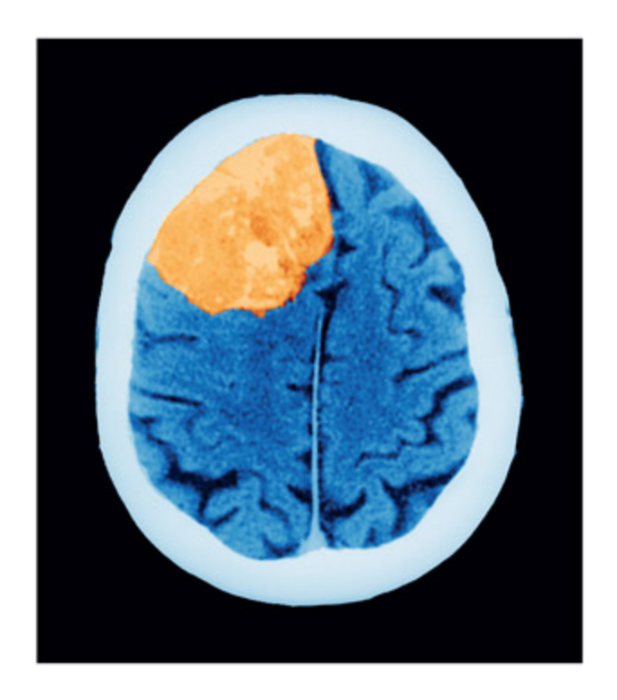
📊 그림 설명
CT 스캔에서 관찰되는 뇌종양의 영상이다. 종양이 주변 뇌 조직을 압박하여 뇌의 형태가 왜곡되어 있으며, 이로 인해 손상 범위를 정확히 추정하기 어렵고 표준 뇌 지도에 매핑하기 곤란함을 보여준다. 이는 종양 환자의 손상-결함 분석에서 구조적 영상 기술의 한계를 시각적으로 예시한다.
A tumor (here shown on a CT scan) can make it hard to estimate lesion size, and the distortion in the shape of the brain makes it hard to map onto a standard atlas. Sovereign, ISM/Science Photo Library.
On finding that a function (F) is disrupted following a lesion to region X, it is tempting to conclude that function F is located in region X or, worse still, that the purpose of region X is to implement F. ese conclusions, and the second one in particular, are tantamount to endorsing a neophrenological view of brain structure–function relationship. Before jumping to su a conclusion, one would need to consider a number of other questions. Is this the only function of region X? Do other regions contribute to the performance of function F, or is this the only region that does so? On finding that a function (F) is disrupted following a lesion to region X, a more cautious conclusion is that region X is critical for performing some aspect of function F. is assertion does not assume that region X has a single function, or that function F has a discrete location. It is also important to
note that even a very discrete brain lesion can disrupt the functioning of distant brain regions that are structurally intact; this is termed diasisis. For example, structural lesions to the le frontal lobe can result in markedly x003C;pg>reduced activity in other distant regions (e.g. le inferior posterior temporal lobe) during a leer judgment task (Price et al., 2001). is can occur even though this distant region is not lesioned and may function normally in other contexts. e implications are that damage to one region can disrupt the functioning of another, intact, region when these two regions work together to implement a particular cognitive function.
Evaluation
Group studies of patients can be important for establishing whether a given region is critical for performing a given task or tasks. Two broad methods are favored, depending on the hypothesis being addressed. e first method involves establishing (on a case-by-case basis) whether a patient is impaired on a given task and then determining the lesion location(s). e second method involves selecting the group on the basis of a lesion to a predefined area and then establishing what functional deficits the group has. is second method is important for testing predictions derived from functional imaging resear.
📊 그림 설명
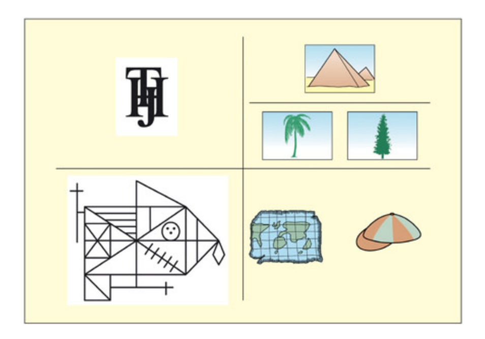
일반적인 신경심리학적 평가 도구들을 보여주는 그림이다. 시계 방향으로 겹쳐진 글자 식별 과제(BORB), 의미 기억 검사(야자수-피라미드 매칭), 실어증 검사(운율 판단, PALPA), 기억 검사(Rey 도형 복사/회상)가 포함된다. 이들은 시각 인식, 의미 기억, 언어, 기억 등 다양한 인지 영역의 손상을 체계적으로 평가하는 데 사용된다.
e purpose of a neuropsyological assessment is to ascertain a patient’s level of functioning relative to that expected based on his or her premorbid functioning (Cipoloi & Warrington, 1995a). Some common neuropsyological tests are shown; clowise from top le: patients with visual recognition problems find it hard to identify overlaid leers relative to non-overlaid ones (from BORB; Riddo & Humphreys, 1995); patients with semantic memory impairments may find it hard to mat the palm tree to the pyramid (Howard & Paerson, 1992); patients with aphasia may find it hard to decide whether things rhyme (from PALPA; Kay et al., 1992); patients with memory problems may be able to copy but not remember this figure
From Rey, 1964. © International Universities Press Inc.
Animal Models in Neuropsyology
Summary
동물 모델은 인간 연구의 한계를 보완하여, 선택적 손상 실험, 단일 세포 기록, 해부학적 연결성 추적 등을 가능하게 한다. 인간과 달리 수술적으로 정밀한 손상을 유도할 수 있고, 동일 개체의 사전/사후 비교가 가능하다. 다만 동물 복지 문제와 언어, 의식 같은 인간 고유 특성의 부재가 한계로 작용한다.
Key Terms
Behavioral neuroscience
비인간 동물 연구의 주요 방법은 단일 세포 기록과 손상 실험이며, 구조적 MRI로 개체별 뇌 해부학적 차이를 보정한다. 실험적 손상 방법에는 흡입, 백질 절단, 신경화학적 손상, 가역적 손상(약물/냉각) 등이 있다. 동물 연구는 일반적으로 행동 신경과학으로 불리며, 영장류 연구에는 엄격한 윤리적 정당화가 요구된다.
Cognitive neuroscience in nonhuman animals.
Summary
비인간 동물 연구에서는 단일 세포 기록과 손상 실험이 주요 방법이며, 구조적 MRI로 전극/손상 위치를 정밀하게 제어한다. 손상 방법으로는 흡입, 백질 절단, 신경화학적 손상, 가역적 손상 등이 있으며, 호스래디시 과산화효소 주입을 통한 해부학적 연결성 추적도 가능하다. 영장류 연구는 동물 복지와 윤리적 정당화가 필수이며, 언어와 의식 같은 인간 고유 특성은 동물 모델의 한계이다.
e two main methods that use non-human animals that are considered in this textbook are single-cell recordings (discussed in Chapter 3) and lesion methods. Both of these methods have been greatly assisted by structural MRI scanning enabling individual differences in ea animal’s brain anatomy to be taken into consideration when placing electrodes and lesions, and also for determining the extent of lesions in vivo. When non-human animals are used in this way, it is typically referred to as behavioral neuroscience rather than cognitive neuroscience. e implication of this difference in terminology is that humans think but animals behave, or, rather, we know that humans think but we can’t be so sure about other animals.
Although lesion methods in humans rely on naturally occurring lesions, it is possible—surgically—to carry out far more selective lesions on other animals. Unlike human lesions, ea animal can serve as its own control by comparing performance before and aer the lesion. It is also common to have control groups of animals that have undergone surgery but received no lesion, or a control group with a lesion in an unrelated area. ere are various methods for producing experimental lesions in animals (Murray & Baxter, 2006):
-
- Aspiration. e earliest methods of lesioning involved aspirating brain regions using a suction device and applying a strong current at the end of an electrode tip to seal the wound. ese methods could potentially damage both gray maer and the underlying white maer that carries information to distant regions.
-
- Transection. is involves cuing of discrete white maer bundles su as the corpus callosum (separating the hemispheres) or the fornix (carrying information from the hippocampus).
-
- Neurochemical lesions. Certain toxins are taken up by selective neurotransmier systems (e.g. for dopamine or serotonin) and, once
-
inside the cell, they create emical reactions that kill it. A more recent approa involves toxins that bind to receptors on the surface of cells, allowing for even more specific targeting of particular neurons.
-
- Reversible “lesions.” Pharmacological manipulations can sometimes produce reversible functional lesions. For example, scopolamine produces a temporary amnesia during the time in whi the drug is active. Cooling of parts of the brain also temporarily suppresses neural activity.
📊 그림 설명
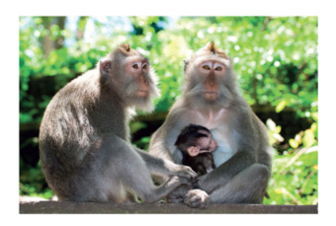
마카크 원숭이 가족의 사진이다. 마카크는 인지 신경과학에서 가장 많이 사용되는 비인간 영장류 중 하나로, 단일 세포 기록과 선택적 손상 실험을 통해 인간 뇌 기능을 연구하는 데 활용된다. 영장류 연구는 엄격한 윤리적 정당화와 동물 복지 기준을 충족해야 한다.
A family of macaque monkeys.
Studies of non-human animals have also enabled a more detailed anatomical understanding of the brain and, in particular, the anatomical connectivity between regions. In non-human animals, injecting the enzyme horseradish peroxidase into axons carries a visible tracer ba to the cell bodies that send them. e tracer can be visualized at post-mortem. is enables one to ascertain whi regions project to a given region (Heimer & Robards, 1981).
While the vast majority of neuroscience resear is conducted on rodents, some resear is still conducted on non-human primates. In many countries, including in the EU, neuropsyological studies of great apes (e.g. impanzees) are not permied. More distant human relatives used in resear include three species of macaque monkeys (rhesus monkey, cynomolgus monkey, and Japanese macaque) and one species of New World
primate, the common marmoset. ere are a number of difficulties associated with the use of animal models in neuropsyology, not least the concern for the welfare of the animals. Scientists working with these species must provide a justification as to why the resear requires primates rather than other animals or other methods, and they must justify the number of animals used. It is also important to have careful breeding programs to avoid having to cat animals in the wild and to protect the animals from viruses. It is important to give them adequate space and social contact. Another disadvantage of animal models is that there are some human traits that do not have obvious counterparts in other species. Language is the most obvious su trait; consciousness is a more controversial one (see Edelman & Seth, 2009).
Transcranial Magnetic Stimulation (TMS)
📊 그림 설명
V5/MT 영역에 TMS를 적용하여 유발된 두 가지 광시(phosphene)의 예시이다. 좌반구 V5/MT 자극은 우측 시야에서 중심에서 멀어지는 방향으로 움직이는 광시를 생성했다. 첫 번째는 “정지된 배경에서 단일 점의 움직임”, 두 번째는 “불연속적으로 우측으로 흐르는” 것으로 보고되었으며, 이는 V5/MT가 운동 지각에 특화된 영역임을 직접적으로 보여준다.
From Stewart et al., 1999. © 1999 Elsevier. Reproduced with permission.
Aempts to stimulate the brain electrically and magnetically have a long history. Electric currents are strongly reduced by the scalp and skull and are therefore more suitable as an invasive tenique on people undergoing surgery. In contrast, magnetic fields do not show this aenuation by the skull. However, the limiting factor in developing this method has been the tenical allenge of producing large magnetic fields, associated with
rapidly anging currents, using a reasonably small stimulator (for a historical overview, see Walsh and Cowey, 1998). Early aempts at magnetic stimulation were successful at eliciting phosphenes (Magnussen & Stevens, 1914), but this was probably due to stimulation of the retina rather than the brain (Barlow et al., 1947). It was not until 1985 that adequate tenology was developed to magnetically stimulate focal regions of the brain (Barker et al., 1985). Since then, the number of publications using this method ology has increased rapidly. Typically, the effects of transcranial magnetic stimu lation (TMS) are small, su that they alter reaction time profiles rather than elicit an overt behavior. But there are instances of the laer. For example, if the coil is placed over the region of the right motor cortex representing the hand, then the subject may experience a sensation or involuntary movement in the le hand (given that the right motor cortex sends movement signals to the le part of the body). If the coil is placed over the right visual cortex, then the subject may report visual sensations or “phosphenes” on the le side (given that the right visual cortex represents the le side of space). Even more specific examples have been documented. Stewart et al., (1999) stimulated a part of the visual cortex dedicated to motion perception (area V5/MT) and reported that these particular phosphenes tended to move. Stimulation in other parts of the visual cortex produces static phosphenes.
How does TMS work?
Summary
TMS는 전자기 유도(Faraday) 원리로 작동하며, 코일의 전류 변화가 자기장을 생성하고, 이 자기장이 뇌 뉴런에 이차 전류를 유도하여 활동전위를 발생시킨다. 8자 코일(figure-of-eight)이 가장 흔하며 자극 초점은 약 1cm²이다. “자기 자극”이라는 명칭은 다소 부정확하며, “전극 없는 비침습적 전기 자극”이 더 정확한 표현이다.
TMS works by virtue of the principle of electromagnetic induction that was first discovered by Miael Faraday. A ange in electric current in a wire (the stimulating coil) generates a magnetic field. e greater the rate of change in electric current, the greater the magnetic field. e magnetic field can then induce a secondary electric current to flow in another wire placed nearby. In the case of TMS, the secondary electric current is induced, not in a metal wire, but in the neurons below the stimulation site. e induced electric current in the neurons is caused by making them “fire” (i.e. generate action potentials) in the same way as they would when responding to
stimuli in the environment. e use of the term “magnetic” is something of a misnomer as the magnetic field acts as a bridge between an electric current in the stimulating coil and the current induced in the brain. Pascual-Leone et al. (1999) suggest that “electrodeless, noninvasive electric stimulation” may be more accurate, although it is a less caty term.
A number of different designs of stimulating coil exist, and the shape of the coil determines how focused the induced current is. One of the most common designs is the figure-of-eight coil. Although the coil itself is quite big, the focal point of stimulation lies at the intersection of the two loops and is about 1 cm2 in area. If you have access to TMS equipment, try holding the coil a few centimeters above your arm. When the pulse is released, you should feel a slight harmless twinge on a small area of skin that is representative of the area of direct stimulation of the brain.
The “virtual lesion”
Summary
TMS는 자극 부위의 뉴런을 인위적으로 활성화하여 진행 중인 인지 활동을 일시적으로 방해하는 “가상 손상” 기법이다. Cohen et al.(1997)의 연구에서 초기 맹인의 후두엽 시각 피질에 TMS를 적용하면 촉각 식별이 손상되었으며, 이는 시각 피질이 촉각 처리에 기능적으로 재편성되었음을 보여준다. TMS는 효과가 짧고 가역적이며, 피험자 내 설계가 가능하고 자극 위치를 자유롭게 이동할 수 있다는 점에서 자연적 손상 방법에 비해 장점이 있다.
TMS causes neurons underneath the stimulation site to be activated. If these neurons are involved in performing a critical cognitive function, then stimulating them artificially will disrupt that function. Although the TMS pulse itself is very brief (less than 1 millisecond), the effects on the cortex may last for several tens of ms. As su, the effects of a single TMS pulse are quily reversed. Although this process is described as a “virtual lesion” or a “reversible lesion,” a more accurate description would be in terms of interference. e neurons are being activated both from an internal source (the task demands themselves) and an external source (the TMS) with the laer disrupting the former. Of course, if the region is not involved in the task, then interference would not occur in this way.
What is the “Visual” Cortex of a Blind Person Used for?
📊 그림 설명
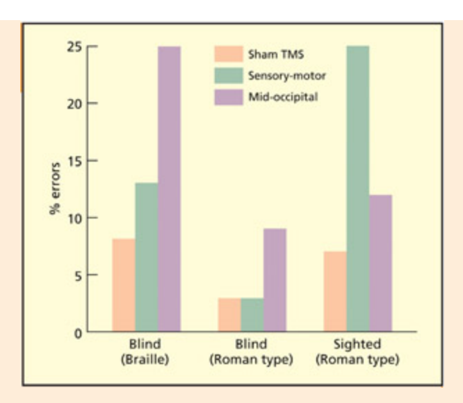
Cohen et al.(1997)의 실험 결과를 보여주는 그래프이다. 초기 맹인에게 중후두엽(시각 피질)에 TMS를 적용하면 촉각 식별이 손상되지만, 눈가린 정상인에게는 영향이 없다. 반면 감각운동 피질 TMS는 정상인의 촉각 식별을 손상시킨다. 이는 초기 맹인의 시각 피질이 촉각 처리로 기능적으로 재편성되었음을 보여주는 핵심 증거이다.
NOTE
Sadato et al. 실험은 fMRI. Cohen이 TMS.
From Cohen et al., 1997. Reprinted by permission of Macmillan Publishers Ltd. © 1997.
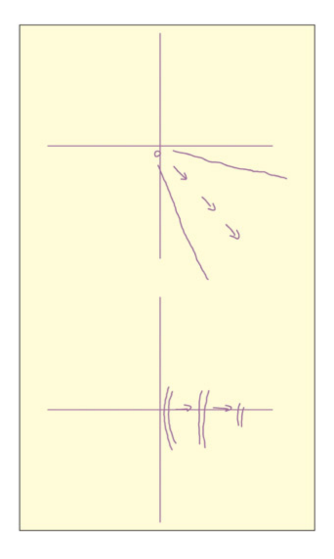
Could whole regions of the brain normally dedicated to one type of processing (e.g. vision) take on a completely different functional aracteristic (e.g. tou)? A number of studies have investigated the functioning of the visual cortex (in the occipital lobes) in people who were blind from a very early age.
Sadato et al. (1996) conducted a brain imaging study demonstrating that early blind Braille readers showed activity in their primary visual cortex (V1) during Braille reading. is was not found for late blind or sighted individuals with their eyes closed. However, functional imaging methods can reveal increases in activity that may not be functionally critical. It could be, for instance, that the blind readers are trying to use the visual cortex during Braille reading but that this activity is not actually contributing to task performance. To address this, lesion methods are appropriate. Given that early blind people with late brain damage restricted to occipital regions are rare (but see Hamilton et al., 2000), TMS avails itself as the most appropriate method.
Cohen et al. (1997) studied tactile identification of Braille leers in early blind individuals, and tactile identification of embossed leers in roman type in both early blind and (blindfolded) sighted individuals. When they placed their finger on the leer, a train of TMS pulses was delivered. e TMS was delivered to a number of sites, including the mid-occipital (“visual” cortex), the sensory-motor (tactile/motor cortex) and “air” as the control condition. For the blind participants, TMS over mid-occipital regions impaired tactile leer discrimination. is suggests that the “visual” cortex is used for tou in the early blind. Sighted people show disruption when TMS is applied over sensory-motor cortex. It is perhaps surprising that blind people do not additionally show an effect here. It could be that, because they are more skilled, they require a higher intensity of TMS for disruption to be induced. ere is evidence for plasticity in somatosensory, as well as mid-occipital, regions in the blind as the region of the brain representing their reading fingers is enlarged by as mu as two or three times (Pascual-Leone & Torres, 1993). Similar TMS studies have revealed cortical enlargements are found for skilled racquet players (Pearce et al., 2000), and cortical reductions found for limb amputees (Cohen et al., 1991). ese suggest that level of use is critical for plasticity.
🧪 Cohen et al. (1997) — 실험 과정과 결과
🎯 연구 질문
초기 맹인의 후두엽(“시각” 피질)이 촉각 처리에 단순히 활성화되는 것을 넘어, 실제로 **필수적(necessary)**으로 기여하는가?
→ Sadato et al.(1996)의 fMRI는 활성화(상관)만 입증할 뿐, 그 활성이 과제 수행에 정말 기여하는지는 알 수 없음 → TMS 가상 손상으로 인과 검증.👥 참가자
- 초기 맹인 (early blind)
- 눈가린 정상인 (blindfolded sighted) — 통제 집단
📋 과제
- 맹인: 점자(Braille) 글자 촉각 식별
- 양 집단 공통: 양각된 로마자(embossed roman letters) 촉각 식별
- 손가락이 글자에 닿는 순간 → TMS 펄스 트레인 전달
📍 TMS 자극 부위 (within-subject 설계)
- 중후두엽 (mid-occipital, “시각” 피질)
- 감각운동 피질 (sensory-motor / tactile-motor cortex)
- 공중(air) — 통제 조건
📊 핵심 결과
자극 부위 초기 맹인 눈가린 정상인 중후두엽 (시각 피질) 촉각 식별 손상 ✅ 영향 없음 감각운동 피질 영향 미미 ⚠️ 촉각 식별 손상 공중 (통제) 영향 없음 영향 없음 ⚠️ 맹인이 감각운동 피질 자극에서 손상을 보이지 않은 점은 의외 → 점자 숙련도가 높아 손상을 유도하려면 더 높은 TMS 강도가 필요했을 가능성(체성감각 영역 자체도 2~3배 확장됨; Pascual-Leone & Torres, 1993).
💡 결론
초기 맹인의 “시각” 피질은 촉각 처리로 **기능적으로 재편성(cortical reorganization)**되었으며, 단순 동반 활성이 아니라 과제 수행에 필수적 역할을 수행한다. fMRI의 상관 한계를 TMS의 가상 손상이 보완한 인과 입증의 대표 사례.
Is it likely that any brain region can substitute for the function of another? In this instance, the function of the brain region is largely the same (i.e. it makes fine-grained spatial discriminations) even though in one instance it responds to vision and in another to tou. However, more recent resear suggests that the occipital cortex in blind individuals can support tasks of a very different nature (e.g. verb generation; Amedi et al., 2004).
📊 그림 설명
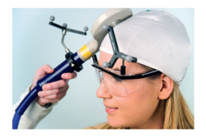
TMS 실험 장면을 보여주는 사진이다. 연구자가 참가자의 두피에 코일을 밀착시키고, 과제 수행 중에 국소적 자기장을 생성하여 특정 뇌 영역을 자극한다. TMS는 코일 위치를 자유롭게 이동할 수 있고 효과가 가역적이어서, 자연적 뇌 손상 연구를 보완하는 비침습적 가상 손상 기법으로 널리 활용된다.
University of Durham/Simon Fraser/Science Photo Library.
임상 사례
Cohen et al.(1997)의 맹인 브레일 연구는 TMS의 임상적 활용을 보여주는 대표적 사례이다. 초기 맹인(early blind)의 후두엽 시각 피질에 TMS를 적용하면 촉각 식별이 손상되었지만, 눈가린 정상인에게는 영향이 없었다. 이는 시각 피질이 촉각 처리로 기능적으로 재편성(cortical reorganization)되었음을 입증한다. 기능적 영상(fMRI)만으로는 “시각 피질의 활성화가 촉각 수행에 실제로 기여하는가?”를 판단할 수 없지만, TMS의 가상 손상 기법으로 그 필수성(necessity)을 확인할 수 있었다.
TMS has a number of advantages over traditional lesion methods (Pascual-Leone et al., 1999). e first advantage is that real brain damage may result in a reorganization of the cognitive system (a violation of the transparency assumption) whereas the effects of TMS are brief and reversible. is also means that within-subject designs (i.e. with and without lesion) are possible in TMS that are very rarely found with organic lesions (neurosurgical interventions are an interest ing exception, but in this instance the brains are not strictly premorbidly “normal” given that surgery is warranted). In TMS, the location of the stimulated site can be removed or moved at will. In organic lesions, the brain injury may be larger than the area under investigation and may affect several cognitive processes.
| • Noreorganization/compensation | • Subcortical lesions can be studied |
|---|---|
| • Can be used to determinetiming of cognition | • Lesions can be accurately localized withMRI (effects of TMS are less well understoodspatially) |
| Advantages of TMS over | Advantages of organic lesions |
|---|---|
| organic lesions | over TMS |
| • Lesion is focal | |
| • Lesion can be moved | • Changes in behavior/cognition are more |
| within the same participant | apparent |
| • Can study functionalintegration |
Will TMS completely replace traditional neuropsyological methods? Probably not. For one thing, TMS is restricted in the sites that can be stimulated, i.e. those beneath the skull; stimulations elsewhere cannot be studied with TMS. Moreover, the spatial extent of the anges induced by TMS is not fully understood and it is possible that more distant brain structures receive stimulation if they are connected to the stimulation site (Paus, 1999). In contrast, organic lesion localization using MRI is more tried and tested. Another advantage of traditional neuropsyology is that the “accidents of nature” turn up some unexpected and bizarre paerns. For example, some patients can name body parts, but not point to named parts of their body (Semenza & Goodglass, 1985); and some patients can draw a bicycle, but not recognize a drawing of a bicycle (Behrmann et al., 1994). Perhaps these sorts of paern could also be observed with TMS, but nobody would think to look for them without the patient-based observations. Indeed, the effects of TMS “lesions” are oen only observable through slowed reaction times and not through error rates or the externally observable behavior that aracterizes most neurological deficits.
Using TMS to study functional integration
Summary
TMS는 기능적 특화(개별 영역의 역할) 외에 기능적 통합(영역 간 상호작용)도 연구할 수 있다. Walsh et al.(1998b)은 V5/MT에 TMS를 적용하여, 운동 정보가 관련된 시각 탐색에서는 수행이 저하되었지만, 운동이 무관한 탐색(녹색 X)에서는 오히려 수행이 향상됨을 발견했다. 이는 서로 다른 시각 영역이 경쟁적으로 작용하며, 무관한 영역을 억제하면 관련 영역의 처리 효율이 증가할 수 있음을 시사한다.
e uses of TMS described so far come within the framework of functional specialization: i.e. trying to understand the functional contributions of particular regions to certain aspects of cognition. A complementary
approa is functional integration; i.e. trying to understand how one region influences another or how one cognitive function influences another. One way in whi this is aieved is by undergoing a session of focal TMS and then studying how this affects the communication between brain regions using fMRI (Bestmann & Feredoes, 2013). (Note: for safety reasons TMS cannot be done in the scanner itself). Another approa is to use TMS to examine competition between brain regions. If there are different processes competing in the brain, then eliminating one process from the competition (using TMS) might have a beneficial effect on the other.
📊 그림 설명
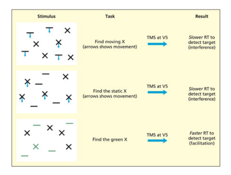
Walsh et al.(1998b)의 시각 탐색 실험 결과를 보여주는 도식이다. V5/MT에 TMS를 적용했을 때, 운동이 과제에 관련된 조건(움직이는 X 찾기)에서는 수행이 저하되었지만, 운동이 무관한 조건(녹색 X 찾기)에서는 오히려 수행이 향상되었다. 이는 서로 다른 시각 영역이 경쟁적으로 작용하며, 무관한 영역의 억제가 관련 영역의 처리 효율을 높일 수 있음을 시사한다.
casacde 식의 정보 전달 과정에서 V5를 망가뜨려, 색상 지각에 의미있는 V4 활동을 가속.
e participants must sear for the presence or absence of a specified target (e.g. moving X) in an array of other items. TMS was applied over area V5/MT (involved in visual motion perception) at various points during sear. If motion was relevant to the sear task, then performance was impaired, but if motion was irrelevant to the sear task, then performance was facilitated. Adapted from Walsh et al., 1998b.
e brain divides up the visual world into different aributes su as color, shape and motion and these different aributes are essentially represented in different regions of the brain (see Chapter 6 for discussion). One theoretical question is: “Do these regions compete with ea other, and
does aending to one aribute (e.g. motion) have positive or negative consequences for irrelevant aributes (e.g. color)?” To answer this question, Walsh et al. (1998b) presented participants with arrays of different shapes made up of different colors that were either moving or static. e task of the participants was to determine whether a prespecified target (e.g. a moving cross, a static cross, a green cross) was present or absent in the array as quily as possible. TMS was delivered at area V5/MT (specialized for visual motion perception) at a number of different time intervals, but, for simplicity, the overall paern across time only will be discussed here. In the first two examples, motion is needed to discriminate between targets and distractors because relying on shape alone will not help (some Xs move and some Xs are static). Unsurprisingly, a virtual lesion to V5/MT disrupts this visual sear, as has been found for organic lesions to this area (McLeod et al., 1989). e unexpected finding comes when there is no motion at all and the participants must find a target based on color and form (a green X). In this instance, a virtual lesion to V5/MT facilitates sear efficiency. is suggests that different visual areas may compete with ea other and eliminating an irrelevant visual area can improve the operation of relevant ones.
Practical aspects of using TMS
Summary
TMS 실험 설계의 핵심 고려사항은 자극 타이밍, 자극 위치, 제어 조건이다. 단일 펄스는 인지 과정의 시간적 정보를 제공하고, 반복 펄스(rTMS)는 영역의 필요성 탐지에 더 효과적이다. 일반적으로 지각 과정 연구는 단일 펄스를, 고차 인지(기억, 언어) 연구는 rTMS를 선호한다.
When designing experiments using TMS (or when evaluating other people’s oice of design), there are three main considerations: when to deliver the pulses, where to deliver the pulses, and selection of appropriate control conditions (for a good overview, see Robertson et al., 2003). Finally, given that the brain is being stimulated, one must be fully aware of safety and ethical considerations when performing TMS experiments.
Timing issues—repetitive or single pulse?
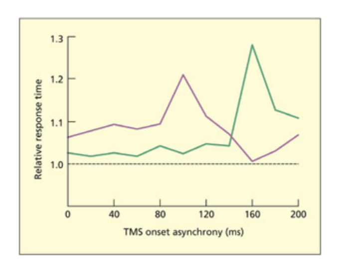
📊 그림 설명
Ashbridge et al.(1997)의 실험에서 TMS의 시간적 해상도를 보여주는 그래프이다. 두정엽에 TMS를 적용했을 때, 목표물 존재 시행은 100ms(보라색 선), 부재 시행은 160ms(녹색 선) 시점에서만 수행이 저하되었다. 이러한 시간적 분리는 비가역적 뇌 손상 환자에서는 관찰할 수 없으며, TMS만의 고유한 장점인 인지 과정의 시간적 특성 탐구를 보여준다.
From Ashbridge et al., 1997. © 1997 Elsevier. Reproduced with permission.
e issue of when to deliver the pulse is crucial to the success, or otherwise, of a TMS experiment. On rare occasions, the time taken for a stimulus to be registered in a given brain region is known by previous resear using other teniques. For example, single-cell recordings suggest that it takes 100 ms for a visual stimulus to be registered in the primary visual cortex (area V1), and TMS studies in whi a single pulse is delivered close to this critical window can render the subject effectively “blind” to the stimulus (Corthout et al., 1999). On most occasions, information su as this will not be known. In this situation, there are a number of options. First, one could make the time of pulse delivery a variable in its own right. For example, if a stimulus is presented for 500 ms, the TMS pulse (or pulses) could be delivered in different time windows (0–50 ms, 50–100 ms, … 450–500 ms). is experimental design could thus provide important information about the timing of cognition, as well as providing information about the necessity of that region. An alternative solution is to use a train of pulses during the task (i.e. repetitive or rTMS). In this situation, the experiment becomes
potentially more powerful in its ability to detect the necessity of a region, but it would not be possible to draw conclusions about timing because it would be unclear whi pulse (or pulses) was critical. Whether or not singlepulse or rTMS is used is not only related to whether timing is an independent variable, but also to the nature of the task itself. Some tasks may require several pulses for TMS to exert interference. e reasons why this might be are not fully understood, but it is a general rule of thumb that TMS studies of perceptual processes have oen used single-pulse designs, whereas studies of “higher” cognition (e.g. memory, language) have oen used rTMS (Walsh & Rushworth, 1999).
How to hit the spot?
Summary
TMS 자극 위치는 두피 랜드마크(inion, nasion, vertex) 기준 좌표나 수영 캡 격자로 정의할 수 있다. 개인별 뇌 해부학적 차이를 반영하기 위해 구조적/기능적 MRI와 결합한 프레임리스 스테레오택시 기법이 사용되며, 인접 위치를 대조 조건으로 활용하여 관심 영역의 공간적 범위를 추정할 수 있다.
To conduct a TMS experiment, one needs to make some assumptions about whi regions of the brain would be interesting to stimulate. In some instances, functional resolution is all that is needed. Just as with the arguments concerning classical versus cognitive neuropsyology, one may wish to establish that a given task/behavior can be selectively disrupted (in whi case, the location of the stimulation site is not relevant to the type of conclusion drawn).
Positions on the head can be defined relative to landmarks, su as those used in the EEG system of electrode placement. Skull landmarks include the inion (a bony protrusion at the ba of the skull), the anion (the bony ridge between the eyebrows), and the vertex (midway between the anion and inion, and midway between the ears). For example, one published way of approximately locating area V5/MT (dedicated to visual motion perception) is by marking a spot 5 cm in front of the inion, and 3 cm up from it (Walsh et al., 1998a). e spot can be physically marked by placing an X on the skin, or by marking the position on a taut swimming cap. If a precise location is not known before the study, then one could stimulate, say, six different spots lying in a 2 · 3 cm grid, drawn on a swimming cap relative to a fixed skull
landmark. Different adjacent positions could then serve as control conditions in the analysis.
Structural and functional MRI can also be used to locate candidate regions of stimulation taking into account individual differences in brain anatomy and skull shape (this is called frameless stereotaxy). A structural or functional MRI scan can be obtained prior to TMS and then online digital registration (using specialist soware) enables the position on the skull to be identified. Alternatively, the TMS could be performed prior to a structural MRI scan in whi the stimulation sites used have been marked in su a way as to render them visible on the scan. Cod liver oil tablets, aaed to the head, have been used previously (Hadland et al., 2001).
What is the appropriate control condition?
Summary
이 섹션에서는 트랜스크란셜 마그네틱 스태뮬레이션(TMS) 실험에서 적절한 제어 조건(control condition)을 설정하기 위한 두 가지 주요 접근법을 제시한다. 첫째, 임계 시간 창(critical time window)과 비임계 시간 창(non-critical time window)에서 동일한 뇌 영역을 자극했을 때의 성능 비교를 통해 기능적 영향을 분석할 수 있으며, 둘째, 임계 영역(critical region)과 비임계 영역(non-critical region) 간의 자극 비교를 통해 뇌 영역의 특이성을 탐구할 수 있다. 비임계 영역 선택 시 접근 영역(adjacent regions)을 활용하면 관심 영역의 공간적 크기를 추가적으로 파악할 수 있고, 인지 기능의 측면화(lateralization)가 명확한 경우 대뇌 반대편의 동일 부위를 대조군으로 사용하는 것이 효과적이다. 또한, 샤ム TMS(Sham TMS)는 코일을 머리에 대지 않고 공중에서 작동시키는 방식이지만, 이는 주변 효과(peripheral effects)를 완전히 제거하지 못해 이상적인 제어 조건이 아니며, TMS 없음(no TMS)을 대조군으로 삼는 것도 바람직하지 않다. 이에 따라 태스크 제어(task control)가 추가적인 방법으로 제안되는데, 이는 동일한 시간과 영역에서 자극을 수행하면서 태스크 요소(예: 자극물, 지시사항)를 변경해 기능적 통합을 분석하는 방식이다.
Two possible control conditions for TMS experiments have already been considered. First, one can compare performance when the same region is stimulated in critical and non-critical time windows. Second, one can compare stimulation in critical and non-critical regions. Some consideration needs to be given to the selection of the non-critical region. Using regions adjacent to the critical region can provide extra clues about the spatial size of the region of interest. In studies in whi there is good reason to believe that the cognitive function is lateralized, one could use the same site on the opposite hemisphere as a control. A further advantage in using the control conditions mentioned above is that peripheral effects of TMS can be minimized. ese include the loud sound of the pulse and twites caused by inadvertent stimulation of the facial nerves and muscles. e laer can be more pronounced at some sites and so using adjacent regions or opposite hemisphere regions would minimize this. “Sham TMS,” in whi the coil is held in the air rather than against the head, is not an ideal control condition, and having no TMS at all as a control condition is also not desirable. Another control condition that can be used in TMS experiments is a task
control. us, the same region can be stimulated at the same times, but with some aspect of the task anged (e.g. the stimuli, the instructions).
Evaluation
TMS is an interesting addition to the cognitive neuroscientist’s toolkit. It is able to ascertain the importance of a given region by stimulating that region during task performance. As su, it is related to other lesion methods that are used for establishing the importance of a given region, but it has certain advantages over the organic lesion method. e main advantage lies in the fact that the interference is short-lived and reversible. It can also be used to explore how regions interact with ea other (functional connectivity) and shed light on the timing of cognitive processes.
주의
TMS를 “자기 자극”이라고 하지만, 실제로 뉴런을 자극하는 것은 자기장이 유도한 전류(electric current)이다. 자기장은 두개골을 통과하는 “다리” 역할만 한다. 또한 TMS의 효과는 피질 표면(cortical surface) 아래의 영역에만 국한되며, 심부 뇌 구조(예: 해마, 기저핵)는 자극할 수 없다. 시험에서 “TMS가 모든 뇌 영역의 기능을 검증할 수 있다”는 진술은 오답이다.
Safety and Ethical Issues in TMS Research
Summary
이 섹션에서는 트랜스크란셜 마그네틱 스태뮬레이션(TMS) 실험에서 고려해야 할 안전성 및 윤리적 문제를 다룬다. 연구자들은 기관별 지역 규정을 준수해야 하며, 특히 **Rossi et al. (2009)**가 제안한 맥스ималь 펄스 수와 강도 기준을 준수해야 한다. 단일 펄스 TMS는 일반적으로 안전하지만, 반복 펄스 TMS는 경련 유발 위험(Wassermann et al., 1996)이 있어 경련성 발작 병력 또는 가족력이 있는 참여자, 심장 기구(pacemaker)나 의료 임플란트 보유자에게는 제외해야 한다. 실험 중 전자기 간섭을 방지하기 위해 카드, 디스크, 컴퓨터 등은 코일에서 1m 이상 떨어진 위치에 두어야 하며, 모터 임계치(motor threshold)에 따라 펄스 강도가 결정된다. 실험 참여자는 100dB 수준의 소음과 면상 근육 수축(facial twitches)으로 인해 불편함을 느낄 수 있어 이어플이나 이어폰 사용이 필수이며, 경련 유발 가능성에 대해 사전에 경고해야 한다. 또한 단일 세션은 장기적 영향이 없지만, 반복 실험은 정신 건강 개선(George et al., 1995)과 같은 긍정적 효과 또는 부작용이 발생할 수 있어, 치료 목적 외에는 짧은 기간 내 동일 참여자 반복 테스트를 피해야 한다.
Researers need to bear in mind a number of safety issues when conducting TMS experiments. It is essential to be aware of the local regulations that apply in your own institution, but the following points are likely to be important:
-
e most recent safety and ethics guidelines come from a consensus of leading researers in the field that offers guidance on issues su as the number and intensity of pulses (Rossi et al., 2009).
-
Whereas single-pulse TMS is generally considered to be safe, repetitive-pulse TMS carries a very small risk of inducing a seizure (Wassermann et al., 1996). Given this risk, participants with epilepsy or a familial history of epilepsy are normally excluded. Participants with pacemakers and medical implants
-
should also be excluded. Credit cards, computer discs and computers should be kept at least 1 m away from the coil.
-
e intensity of the pulses that can be delivered is normally specified with respect to the “motor threshold”; that is, the intensity of the pulse, delivered over the motor cortex, that produces a just noticeable motor response (for a discussion of problems with this, see Robertson et al., 2003).
-
During the experiment, some participants might experience minor discomfort due to the sound of the pulses and facial twites. Although ea TMS pulse is loud (~100 dB), the duration of ea pulse is brief (1 ms). Nonetheless, it is mandatory to protect the ears with earplugs or headphones. When the coil is in certain positions, the facial nerves (as well as the brain) may be stimulated, resulting in involuntary twites (e.g. blinking, jaw clamping). Participants should be warned of this and told they can exercise their right to withdraw from the study if it causes too mu discomfort.
-
It is generally believed that a single session of TMS has no long-term consequences. However, repeated participation in experiments could conceivably have longer-term effects—either positive or deleterious. A number of studies report an improvement in mood in depressed individuals following repeated frontal lobe stimulation (George et al., 1995). But this study involved repeated stimulation on a daily basis. Except in cases of therapeutic intervention, it is good practice not to test the same participants many times over a short interval.
Transcranial Direct Current Stimulation (tDCS)
Summary
이 섹션에서는 트랜스크란셜 디렉트 커런트 스태뮬레이션(tDCS)이라는 비침습적 뇌 자극 기법을 소개하며, 이는 약한 전류를 두피에 적용하여 신경 활동을 조절하는 방식으로 작동한다고 설명한다. TMS(트랜스크란셜 마그네틱 스태뮬레이션)와 유사한 가상 손상(virtual lesion) 접근법을 기반으로 하되, 전기 자극을 활용해 인지 기능의 일시적 손상(temporarily disrupt cognitive function)뿐만 아니라 인지 기능 향상(boost cognitive function)도 가능하다는 점에서 차별화된다. 특히, 전극을 두피에 배치하여 특정 뇌 영역을 자극함으로써 신경회로 기능 조절이나 인지/운동 기능 분석에 활용되며, 스위밍 캡에 그려진 격자(2×3 cm 크기)를 사용해 다중 위치 자극이 가능하다는 실용적 고려사항도 제시된다. 이 기법은 뇌 손상 환자 분석이나 그룹 연구와는 달리 직접적인 신경 활성 조절을 가능하게 하며, 인지 신경과학에서 기능적 통합(functional integration) 연구에 기여할 수 있는 잠재력을 보유하고 있다.
Key Terms
Cathodal tDCS
Decreases cortical excitability and decreases performance.
카토달 tDCS는 피질 흥분성을 감소시키고 인지/운동 성능 저하를 유발한다. 가상 손상(virtual lesion)과 유사한 원리로, 전기 자극이 뉴런 활동을 억제하여 기능적 손상을 일시적으로 유도한다.
Anodal tDCS
Increases cortical excitability and increases performance.
아노달 tDCS는 피질 흥분성을 증가시키고 인지 성능을 향상시킨다. 시각 피질에 아노달 자극을 가하면 초기 시각 ERP 성분(N100) 진폭이 증가하고, GABA 시스템에 영향을 미친다. 즉각적 효과는 휴식 상태 막 잠재력에, 지속 효과는 시냅스 가소성 변화를 통해 학습과 신경전달물질 시스템에 작용한다. 뇌졸중 환자의 운동 기능 회복, 파킨슨병 환자의 작업 기억 향상 등 재활 치료에서도 효과가 확인되었다.
e use of electrical currents to stimulate the brain has a long and eered history, with the most notorious noninvasive method being electroconvulsive therapy (ECT) used to “treat” psyiatric illnesses. Unlike ECT, the method of transcranial direct current stimulation (tDCS) uses a very weak electric current. Direct current involves the flow of electric arge from a positive site (an anode) to a negative site (a cathode). In tDCS, a stimulating pad (either anodal or cathodal) is placed over the region of interest and the control pad is placed in a site of no interest (sometimes on the front of the forehead, or sometimes on a distant site su as the shoulders). Aer a period of stimulation (e.g. 10 min) a cognitive task is performed and this can be compared with sham stimulation, or anodal and cathodal stimulation can be directly contrasted. Cathodal tDCS stimulation tends to disrupt performance (i.e. it is conceptually equivalent to a virtual lesion approa) whereas anodal tDCS stimulation tends to enhance performance (Nitse et al., 2008). For example, anodal stimulation over visual cortex leads to an enhanced early visual ERP component (N100) and enhances the ability to detect weak visual stimuli, whereas cathodal
stimulation has the opposite effects (Accornero et al., 2007; Antal et al., 2001).
Stagg and Nitse (2011) provide a summary of the likely neurophysiological meanisms. It is important to consider the immediate effects of direct current stimulation and the aereffects separately. Animal models of direct current stimulation followed by single-cell recordings have shown that anodal stimulation increases the spontaneous firing rate of neurons whereas cathodal stimulation reduces the firing rate. e immediate effects of stimulation are believed to occur on the resting membrane potential rather than modulation at the synapse. However, the aftereffects of stimulation are likely to occur due to anges in synaptic plasticity influencing learning and perhaps affecting different neurotransmier systems. Anodal stimulation affects the GABA system (this neurotransmier has inhibitory effects) whereas cathodal stimulation affects the glutamate system (this neurotransmier has excitatory effects).
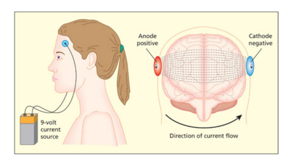
📊 그림 설명
tDCS 장치의 구성과 작동 원리를 보여주는 도식이다. 두피에 부착된 자극 패드를 통해 약한 직류 전류가 양극(anode)에서 음극(cathode)으로 흐른다. 아노달 자극은 피질 흥분성을 증가시키고, 카토달 자극은 감소시키며, 이 양방향 조절이 tDCS를 인지 기능 향상과 가상 손상 연구 모두에 활용할 수 있게 한다.
Adapted from George and Aston-Jones.
e current safety guidelines recommend upper limits on the size of the current and the surface area of the stimulating electrodes (Nitse et al.,
2003). If the current is concentrated on a small electrode, then it can cause skin irritation. However, unlike TMS, participants oen cannot tell whether the maine is swited on or used as sham (there is no sound or twiting). As su there is very lile discomfort.
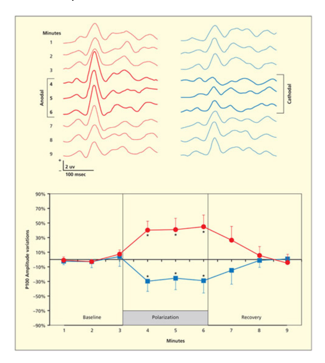
📊 그림 설명
시각 피질에 3분간 tDCS를 적용한 후 흑백 체커보드 자극에 대한 ERP 성분(P100) 진폭 변화를 보여주는 그래프이다. 아노달 자극은 P100 진폭을 증가시키고, 카토달 자극은 감소시켰다. 이는 tDCS가 피질 흥분성을 양방향으로 조절할 수 있음을 전기생리학적으로 입증하는 핵심 데이터이다.
From Accornero et al., 2007.
임상 사례
tDCS의 양방향 조절 능력은 재활 치료에서 큰 잠재력을 보여준다. 아노달 tDCS(피질 흥분성 증가)는 뇌졸중 후 운동 장애 환자의 손 기능 회복, 실어증 환자의 언어 능력 향상, 파킨슨병 환자의 작업 기억 개선에 효과가 입증되었다. 반면 카토달 tDCS(피질 흥분성 감소)는 가상 손상 효과로 연구 목적에 활용된다. TMS와 달리 tDCS는 소음이나 안면 근육 수축이 거의 없어 참가자가 자극 여부를 구분하기 어려워, 이중 맹검(double-blind) 실험 설계에 특히 유리하다.
Repeated sessions of anodal tDCS are becoming increasingly used for cognitive enhancement (of normal brains) and neurorehabilitation (of damaged brains). For instance, repeated sessions of tDCS over the primary motor cortex leads to increased cortical excitability and greater hand functionality in patients with motor impairments following stroke (Hummel et al., 2005). In this study, the treatment was compared with sham and the procedure was double blind (i.e. neither participant nor experimenter knew whi condition they were in). Other studies using anodal tDCS have reported improvements in language following stroke (Monti et al., 2008) and improved working memory in patients with Parkinson’s disease (Boggio et al., 2006).
Summary and Key Points of the Chapter
Summary
이 섹션에서는 이중 분리(double dissociation) 현상을 통해 두 환자가 서로 다른 작업에서 인지 기능의 유의미한 차이를 보일 때, 해당 작업이 서로 다른 신경 자원(neural resources)에 의존함을 설명한다. 또한 단일 사례 연구(single-case studies)는 인지 구성 요소의 조직 방식과 분리 가능성을 이해하는 데 중요한 통찰을 제공하며, 그룹 연구(group studies)는 뇌 손상 위치와 행동 결함 간의 연관성을 규명하고, 기능적 영상 데이터와의 일관성을 검증하는 데 핵심적인 역할을 한다. 트랜스크란셜 마그네틱 스태뮬레이션(TMS)은 코일을 통해 뇌 피질을 자극하여 특정 영역의 기능을 일시적으로 방해함으로써 해당 영역의 작업 수행에 필수적인 역할을 밝혀내는 “가상 손상”(virtual lesion) 기법으로 작동한다. 반면, 트랜스크란셜 디렉트 커런트 스태뮬레이션(tDCS)은 TMS보다 시간 및 공간 해상도는 낮지만, 아노달(anodal) 자극을 통해 인지 기능을 향상시킬 수 있는 장점을 가진다. 이 두 기법은 뇌 기능의 역할을 탐구하는 데 서로 보완적인 접근법으로 활용된다.
- A double dissociation between two patients occurs when patient 1 is significantly beer than patient 2 on task A, and patient 2 is significantly beer than patient 1 on task B. e standard interpretation of this is that task A and task B utilize some different neural resources.
- e use of single cases has led to important insights into the way in whi cognitive components are organized and may be fractionated.
- Group studies of patients are important for making links between lesion location and behavioral deficits, and provide an important source of converging evidence for functional imaging data.
- Transcranial magnetic stimulation (TMS) works by stimulating a region of cortex placed beneath a current-carrying coil. is stimulation temporarily interferes with ongoing cognitive activity in that region and, therefore, provides information about the necessity of that region for performing the task. is has been termed a “virtual lesion.”
Transcranial direct current stimulation (tDCS) has a poorer temporal and spatial resolution to TMS, but has the advantage of being able to facilitate cognitive function (anodal tDCS).
Example Essay Questions
Summary
이 섹션에서는 뇌 손상 환자의 인지 기능 분석을 통해 정상 인지 기능을 이해하는 데 필요한 가정을 비판적으로 평가하며, 특히 그룹 연구(group studies)와 단일 사례 연구(single-case studies)의 역할을 논의한다. 그룹 연구는 환자 간 변이성을 통계적으로 평균화해 집단 수준의 인지 패턴을 도출하지만, 개별 차이나 신경 가소성(neural plasticity)을 무시할 수 있는 한계가 있다. 반면 단일 사례 연구는 특정 환자의 복잡한 인지 기능 장애를 세부적으로 탐구할 수 있지만, 일반화 가능성이 제한된다. TMS(트랜스크란셜 마그네틱 스태뮬레이션)와 tDCS(트랜스크란셜 디렉트 커런트 스태뮬레이션)는 뇌 기능의 일시적 조절을 통해 신경 회로의 유연성(plasticity)을 실증적으로 탐구하는 데 기여했으며, 이는 자연적인 뇌 손상(organic lesion)과의 비교를 통해 인위적 자극 방법의 잠재력을 강조한다. 특히 TMS/tDCS는 기능적 통합(functional integration) 및 흥분성 조절(modulation of excitability)을 통해 뇌 가소성의 동적 메커니즘을 규명하는 데 중요한 역할을 하며, 이는 역공학적 접근(reverse engineering)과 연계된 인지 신경과학(cognitive neuroscience)의 핵심 주제로 작용한다. 그러나 뇌 손상 기반 연구와 자극 기법 간의 상호 보완성과 한계를 명확히 인식하는 것이 중요하다.
- What assumptions must one accept to be able to draw inferences about normal cognition from adults with brain damage? Are these assumptions plausible?
- Critically evaluate the role of group studies in neuropsyological resear.
- What are the advantages and disadvantages of using single cases to draw inferences about normal cognitive functioning?
- How have TMS and tDCS studies contributed to our knowledge of brain plasticity?
- Compare and contrast lesion methods arising from organic brain damage with TMS and tDCS.

📊 그림 설명
교재의 동반 웹사이트를 안내하는 그림이다. 웹사이트에서는 핵심 논문 참고문헌, 주요 주제에 대한 비디오 강의 및 인터뷰, 다중 선택 문제와 대화형 플래시카드, 다운로드 가능한 용어집 등 학습 보조 자료를 제공한다.
Visit the companion website at www.psypress/cw/ward for:
- References to key papers and readings
- Video lectures and interviews on key topics with leading psyologists Elizabeth Warrington and author Jamie Ward, as well as demonstrations of and lectures on brain stimulation
- Multiple oice questions and interactive flashcards to test your knowledge
Downloadable glossary
Recommended Further Reading
Summary
이 섹션에서는 신경심리학 분야에서 단일 사례 연구(single-case studies)와 그룹 연구(group studies)의 방법론적 문제를 다룬 Cognitive Neuropsychology(1988, Vol. 5, No. 5) 특집호 및 Cortex(2003, 39(1))의 기능적 분리(dissociations) 활용에 대한 논쟁을 소개한다. 또한 트랜스크란셜 마그네틱 스태뮬레이션(TMS)에 대한 기술적 설명과 임상 적용 사례를 중심으로 한 Pascual-Leone 등(2002)의 Handbook of transcranial magnetic stimulation과, 인지 신경과학(cognitive neuroscience) 관점에서 TMS의 메커니즘을 분석한 Walsh 및 Pascual-Leone(2003)의 Transcranial magnetic stimulation: A neurochronometrics of mind을 추천 자료로 제시한다. 특히 이 두 저서는 TMS의 임상(clinical)과 인지 기능 분석(cognitive neuroscience)에 대한 차별화된 접근법을 설명하며, 이전 섹션에서 논의된 뇌 자극 기법(TMS, tDCS)의 이론적 기반과 실험 설계에 대한 심층적 이해를 돕는 자료로 기능한다.
- A special edition of Cognitive Neuropsychology (1988), Vol. 5, No. 5, is dedicated to methodological issues related to singlecase and group studies in neuropsyology.
- A special section of the journal Cortex (2003), 39(1), debates the use of dissociations in neuropsyology.
- Pascual-Leone, A., Davey, N. J., Rothwell, J., Wassermann, E. M., & Puri, B. K. (2002). Handbook of transcranial magnetic stimulation. London: Arnold. A detailed account of the methods and uses of TMS, with a strong clinical slant.
- Walsh, V. & Pascual-Leone, A. (2003). Transcranial magnetic stimulation: A neurochronometrics of mind. Cambridge, MA: MIT Press. A detailed account of the methods and uses of TMS, with a cognitive neuroscience slant.